Case 1
25 years old women with a palpable left breast mass,
presented as focal acoustic shadowing without a mass configuration on ultrasound.
Mammography was negative because of dense breast tissue.
The patient was diagnosed as sclerosing adenosis by core needle biopsy under ultrasound (Fig1).
Sclerosing adenosis is a benign proliferative disease of the breast.
On clinical,
radiological,
and even histopathological examination,
it can be confused with malignancy.
On histopathological examination,
sclerosing adenosis is present in 12% of benign and 5%–7% of malignant specimens (1).
The disease has an increased incidence among reproductive-age and perimenopausal women,
especially between 35 and 50 years of age (1–4),
while our patient with sclerosing adenosis was 25 years old.
There is no typical radiological criterion for diagnosis.
The characteristic findings of sclerosing adenosis on mammography and ultrasonography have only rarely been described in the literatüre (5–6).
On mammographic imaging,
in asymptomatic patients,
various findings such as mass lesions,
microcalcifications,
focal asymmetrical opacities and architectural distortions can be detected.
If there is a palpable mass,
a tumoral mass lesion can also be detected by both mammography and ultrasonography. Our patient had palpable mass in the left breast with normal mammogram because of dense breast tissue, presented with only focal acoustic shadowing on ultrasound.
Core needle biopsy or excisional biopsy can be used for diagnosis.
In the diagnosis of sclerosing adenosis,
core needle biopsy can be the first step (1).
Our patient was diagnosed by core needle biopsy under ultrasound.
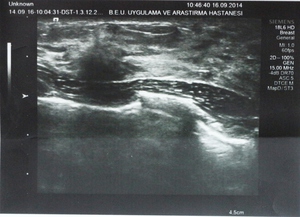
Fig. 1: 25 years old women with a palpable left breast mass, presented as focal acoustic shadowing without a mass configuration on ultrasound.
References: Bulent Ecevit University, School Of Medicine, Department of Radiology,Zonguldak, Turkey
Figure 1. 25 years old women with a palpable left breast mass,
presented as focal acoustic shadowing without a mass configuration on ultrasound.
Case 2:
The women with a palpable right breast mass,
showed encapsulated masses with a heterogeneous appearance with smooth lobulations on US.
Mammography was negative because of dense breast tissue (Fig 2).
Breast hamartoma is an uncommon breast tumor that accounts for approximately 4.8% of all benign breast masses.
Breast hamartomas were initially defined as well-circumscribed tumors composed of varying amounts of epithelial elements in a fibrofatty stroma (7).
Although different studies have used different criteria,
the most common histologic feature is the presence of lobules within a fibrotic stroma (8).
Hamartomas have a mammographic appearance typical of lucent lesions containing fat,varying radiodense fibrous and adenomatous elements,
sharp margins,
and,
in some cases,
a thin capsule (9).
Conversely,
breast hamartomas have wide sonographic variability (7-9).
Our case had normal mammogram while showed encapsulated mass with a heterogeneous appearance on US.
Our patient had core needle biopsy under ultrasound and was diagnosed fibroadenomatoid mass containing fat tissue (Figure 2-4).
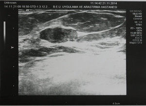
Fig. 2: The women with a palpable right breast mass, showed encapsulated masses with a heterogeneous appearance with smooth lobulations on US.
References: Bulent Ecevit University, School Of Medicine, Department of Radiology,Zonguldak, Turkey
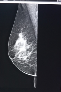
Fig. 3: Mammography was negative because of dense breast tissue.
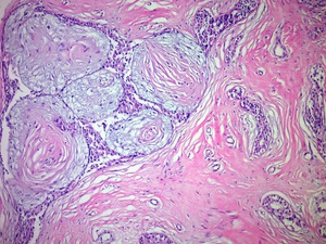
Fig. 4: Morphological appearance compatible with fibroadenomatoid changes. The final diagnosis was hamartoma.
References: Bulent Ecevit University, School Of Medicine, Department of Pathology,Zonguldak, Turkey
Figure 2.The women with a palpable right breast mass,
showed encapsulated masses with a heterogeneous appearance with smooth lobulations on US.
Figure 3. Mammography was negative because of dense breast tissue.
Figure 4. Morphological appearance compatible with fibroadenomatoid changes.
The final diagnosis was hamartoma.
Case 3
45 years old women with a palpable left breast mass,
who had a mass lesion on mammogram while circumscribed mass with smooth lobulations,
slightly hyperechoic relative to the normal fat lobules,
but was not as echogenic as the adjacent fibroglandular tissue and diagnosed by core needle biopsy under ultrasound (Figure 5-7).
Angiolipoma is benign neoplasm,
described as an uncommon variant of lipoma with vascular proliferation among mature adipocytes.
While typically occuring on the trunk and extremities as multiple painful nodules in the subcutaneous tissues,
they rarely occur in the breast (10).
Angiolipoma has nonspecific imaging features.
The most common mammographic appearance of angiolipomas of the breast in this series was an oval or round,
isodense,
circumscribed mass.
The most common sonographic features were oval shape,
circumscribed borders,
and iso- to slight hyperechogenicity.
Biopsy is required to ultimately make this diagnosis.
Similarly,
our case had a isodense,
circumscribed mass lesion on mammogram while echogenic mass which diagnosed by core needle biopsy under ultrasound (Figure 5-7).
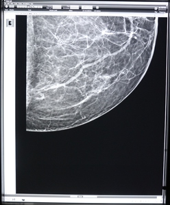
Fig. 5: 45 years old women with a palpable left breast mass, who had a circumscribed isodense mass lesion on mammogram
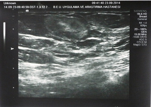
Fig. 6: The mass has smooth lobulations, slightly hyperechoic relative to the normal fat lobules on sonogram.
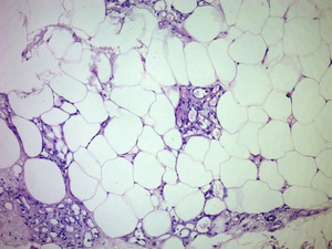
Fig. 7: Lipomatous tissue rich in vascular structures some of which contains thrombus. The final diagnosis was angiolipoma
References: Bulent Ecevit University, School Of Medicine, Department of Pathology,Zonguldak, Turkey
Figure 5. 45 years old women with a palpable left breast mass,
who had a circumscribed isodense mass lesion on mammogram.
Figure 6. The mass has smooth lobulations,
slightly hyperechoic relative to the normal fat lobules on sonogram.
Figure 7. Lipomatous tissue rich in vascular structures some of which contains thrombus.
The final diagnosis was angiolipoma.
