ECR 2015 / C-1725
Novel interventions of the Achilles tendon and related structures
This poster is published under an open license. Please read the disclaimer for further details.
Congress:
ECR 2015
Poster Number:
C-1725
Type:
Educational Exhibit
Keywords:
Musculoskeletal bone, Musculoskeletal joint, Musculoskeletal system, MR, Ultrasound, Ultrasound-Power Doppler, Contrast agent-intravenous, Arthritides, Athletic injuries
Authors:
A. N. Tavare, S. Tincey, H. Madani, O. Chan; London/UK
DOI:
10.1594/ecr2015/C-1725
. The tendon is of uniform thickness and the parallel fibrillar structure is clearly seen. References: otto chan")
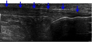
Fig. 1:
Longitudinal ultrasound shows the normal Achilles tendon (blue arrows). The...
 which is has a normal flat contour and with fibres clearly visible")
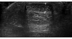
Fig. 2:
Axial ultrasound shows normal tendon (between calipers) which is has a normal...
")
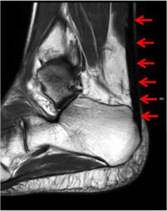
Fig. 3:
Sagittal T2-weighted MRI illustrates the normal Achilles tendon (red arrows)
 References: otto chan")
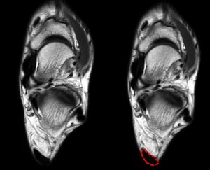
Fig. 4:
Axial T2-weighted MRI shows the normal Achilles tendon (red line)

Fig. 5:
Diffuse tendon enlargement with hypoechoic areas and disruption of normal...

Fig. 6:
Power Doppler demonstrates diffuse abnormal hypervascularity of the...
, which is also enlarged, near the calcaneum.")
Fig. 7:
Sagittal fat-supressed proton density MRI shows increased signal within the...
 and the tendinopathic insertional component (blue arrow) near the calcaneum (C) with ill-defined hypoechoic change and loss of normal fibrillary pattern.")
Fig. 8:
Longitudinal ultrasound shows normal midsubstance Achilles tendon (AT) and the...

Fig. 9:
Longitudinal ultrasound of a different patient again shows similar...

Fig. 22:
Axial schematic shows the relationship of the FC to the paratenon and Achilles...
 encircling the Achilles tendon;
B - motion of fascia cruris during inversion and eversion of the ankle and the site of injury (red) when the fascia cruris tears during powerful movements of the ankle. Injuries may either occur on the medial or lateral sides. References: otto chan")
Fig. 23:
Axial MRI sections demonstrate:
A - the fascia cruris (FC) encircling the...
 clearly distinct from the Achilles tendon (A), consistent with FC tear. References: otto chan")
Fig. 24:
Oblique axial ultrasound shows a large globular hypoechoic area in the lateral...
 is clearly seen to be distinct from the FC tear. The normal thin FC is seen on the anterolateral side of the Achilles tendon. References: otto chan")
Fig. 25:
Axial T2-weighted MRI shows globular area of intermediate signal within medial...