ECR 2015 / C-1911

Cervical Thymic Anomalies: A Pictorial Review
This poster is published under an open license. Please read the disclaimer for further details.
Congress:
ECR 2015
Poster Number:
C-1911
Type:
Educational Exhibit
Keywords:
Congenital, Normal variants, eLearning, Ultrasound, MR, CT, Head and neck
Authors:
S. M. Tochetto, O. C. Saito, J. D. Zavariz, M. C. Chammas; São Paulo/BR
DOI:
10.1594/ecr2015/C-1911
 195:1-15) References: Sandra Monica Tochetto")
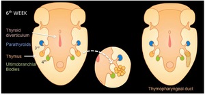
Fig. 1:
Fig 1.: Embryological development of the thymus
(Images modified from Manley,...
 195:1-15) References: Sandra Monica Tochetto")
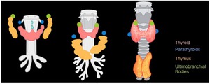
Fig. 2:
Fig 2.: Embryological development of the thymus
(Images modified from Manley,...