ECR 2015 / C-2089
Ultrasound of the elbow joint - anatomical review of normal structures
This poster is published under an open license. Please read the disclaimer for further details.
Congress:
ECR 2015
Poster Number:
C-2089
Type:
Educational Exhibit
Keywords:
Anatomy, Musculoskeletal system, Musculoskeletal joint, Ultrasound, Comparative studies, Technical aspects, Trauma
Authors:
D. Castelo1, E. Matos2, F. C. Pires1; 1Vila Nova de Gaia/PT, 2Porto/PT
DOI:
10.1594/ecr2015/C-2089
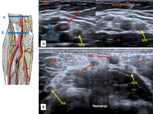
; Bt - Bicipital tendon; BA - Brachial artery; VC - "vena comitants" of brachial artery (deep brachial veins); MN - Median nerve; RN - Radial nerve.]")
Fig. 9:
Anterior elbow, transverse plane:
Image A - veins collapse with compression,...

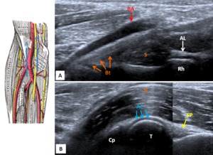
Fig. 10:
Anterior elbow, longitudinal plane:
Image A - Bicipital tendon inserting at...

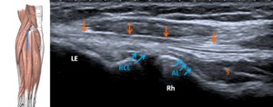
Fig. 11:
Lateral elbow, longitudinal plane:
Vertical arrows - common extensor tendon;...
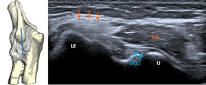
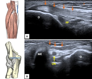
; UCL - ulnar collateral ligament; ME - Medial epicondyle; U - ulna")
Fig. 12:
Medial elbow, longitudinal plane:
Vertical arrows - common flexor tendon; Fm -...

Fig. 13:
Posterior elbow. Image A - longitudinal plane showing the tricipital tendon's...