Anatomy of the elbow
1.
The elbow joint
The elbow joint is one of the most complex in the human skeleton,
actually it comprises two joints that act together,
the humero-ulnar and the humero-radial,
the first articulating the humeral trochlea with the ulnar trochlear notch,
and the second,
the humeral capitulum with the radial head.
Distal to these,
there is the proximal radioulnar joint,
composed by the radial head and the radial notch of the ulna.
All of these joints are covered by the same synovial membrane,
forming a communicating articular cavity.
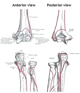
Fig. 1: Bones of the elbow joint - Adapted from Gray's Anatomy 20th edition (1918) with original labels
(some nomenclature may not be up to date)
References: Public Domain (PD)
1.1.
Joint classification and function
As synovial articulations,they can be further classified,
the humero-radio-ulnar joint being a hinge joint and the radio-ulnar an uniaxial pivot joint.
They allow flexion and extension of the forearm (humero-radio-ulnar joint) and are partially responsible for the pronation and supination of the forearm (proximal radio-ulnar joint).
1.2.
Ligamentous structures
Despite the apparent lack of congruence,
the elbow joint is one of the most stable in the human skeleton.
This stability is maintained mainly by medial and lateral collateral ligaments and the fibrous joint capsule.
- Ulnar collateral ligament (UCL) - possesses a triangular shape with it's apex centered at the medial epicondyle and base attached to the medial surface of the proximal ulna.
It is composed by three thick parts united by a thinner wider band.
The anterior part is the strongest and connects the medial epicondyle to the lateral surface of the coronoid process; the posterior part inserts slightly posteriorly at the medial epicondyle and attaches to the near olecranon surface.
The third part,
the oblique,
bridges the notch formed between the medial surface of the olecranon and the coronoid process; from the medial epicondyle,
between the anterior and posterior parts,
thinner fibers descend to meet the oblique part.
The anterior part maintains it's tension during the majority of the movement range and the posterior part gains maximum tension from middle to full flexion.
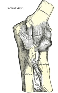
Fig. 2: Ulnar collateral ligament - Adapted from Gray's Anatomy 20th edition (1918) with original labels
(some nomenclature may not be up to date)
References: Public Domain (PD)
- Radial collateral ligament (RCL) - similarly to the UCL,
also defines a triangular shape,
and has it's apex inserted at the infero-lateral portion of the lateral epicondyle.
The base of this triangle inserts in a different fashion,
the majority of fibers attach to the annular ligament with only a minority of it’s posterior fibers inserting at the supinator crest of the ulna. Another ligament acts to stabilize the lateral portion of the elbow,
the lateral ulnar collateral ligament (LUCL); it also has it’s origin at the lateral epicondyle but has it’s insertion slightly posterior to the annular ligament; the LUCL helps supporting the radial head,
preventing it’s posterior dislocation.The RCL is tense along the majority of movement amplitude.
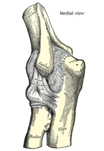
Fig. 3: Radial collateral ligament - Adapted from Gray's Anatomy 20th edition (1918) with original labels
(some nomenclature may not be up to date)
References: Public Domain (PD)
1.3.
Periarticular structures
Other relevant structures in intimate relation with the joint are fat pads and bursae.
The main fat pads lay between the capsule and the synovial membrane and are located at the ulnar,
radial and olecranon fossae of the humerus.
The anterior pads are compressed by the brachialis muscle at full extension and the fat pad at the olecranon fossa is compressed by the triceps during flexion of the joint.
There are many bursae adjacent to the elbow,
the most constant and important is the olecranon bursa,
located in the subcutaneous tissue lining the superior and posterior surface of the olecranon.
The main function of these ancillary structures is to help protect and dampen from trauma.
2.
Muscles crossing the elbow joint
The muscles of this region are usually divided in four groups,
the anterior,
lateral,
medial and posterior groups.
2.1.
Anterior group
- Biceps brachii - the most superficial of this group,
has two heads at it's origin,
the long head from the supraglenoid tubercle and the short head from the coracoid process; the two unite 6 to 7 cm proximal to the elbow and form a common tendon which inserts at the radial tuberosity posteriorly; from this tendon,
originates the thinner bicipital aponeurosis which fuses with the fascia of the flexor muscles.
- Brachialis - has a broad origin from the distal anterior surface of the humerus and from both brachial intermuscular septa,
travels distally,
superfícial to the elbow's capsule and inserts into the coronoid process.
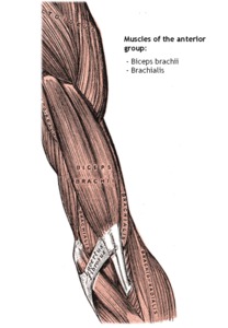
Fig. 4: Left upper arm muscles - Adapted from Gray's Anatomy 20th edition (1918) with original labels
(some nomenclature may not be up to date)
References: Public Domain (PD)
2.2.
Lateral group
- Brachioradialis - has it’s origin proximally to the lateral epicondyle,
at the supracondylar ridge and at the lateral intermuscular septum.
It then crosses the elbow assuming an antero-lateral position.
It inserts proximally to the radial styloid process.
- Extensor muscles of the wrist - in this sub-group are included five muscles,
the extensor carpi radialis longus (ECRL),
extensor carpi radialis brevis(ECRB),
extensor digitorum (ED),
extensor digiti minimi (EDM) and extensor carpi ulnaris (ECU).
The ECRL origin is located inferiorly to the brachioradialis muscle,
also at the lateral supracondylar ridge and lateral intermuscular septum,
to then descend deeply to the brachioradialis.
The remaining extensors (ECRB,
ED,
EDM and the humeral head of the ECU) have a common origin,
at the lateral epicondyle,
and at the elbow level appear as a single muscle that travels distally from lateral to posterior aspect of the forearm.
- Supinator - the deepest of this group,
has two heads,
the superficial head that originates at the posterior aspect of the lateral epicondyle and a deep head that originates at the supinator crest of the ulna.
At it’s origins some fibers attach to the radial collateral and annular ligaments.
This muscle embraces the proximal extremity of the radius,
inserting on the lateral radial surface at this level.
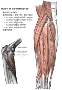
Fig. 5: Right elbow and forearm showing the muscles of the lateral group - Adapted from Gray's Anatomy 20th edition (1918) with original labels
(some nomenclature may not be up to date)
References: Public Domain (PD)
2.3.
Medial group
- Pronator teres - the most superficial of this group,
has two heads,
the humeral head that originates proximally to the medial epicondyle and an ulnar head at the coronoid process.
Travels from medial to lateral along the forearm to insert at the radial shaft.
- Flexors of the hand and wrist - in this sub-group are included six muscles,
the flexor carpi radialis (FCR),
palmaris longus (PL),
flexor carpi ulnaris(FCU),
flexor digitorum superficialis (FDS),
flexor digitorum profundus (FDP) and the flexor pollicis longus (FPL).
The first four (FCR,
PL,
FCU and FDS),
have a common tendon that originates from the medial epicondyle.
They then travel along the anterior aspect of the forearm until they reach the wrist and hand.
The FCU also has an ulnar head that parallels the humeral head and has it’s origin at the medial aspect of the coronoid process.
The FDP and the FPL have their origins at the proximal extremities of the ulna and radius (respectively) and distally insert at their corresponding digits (the FPL at the first digit and the FDP at the remaining).
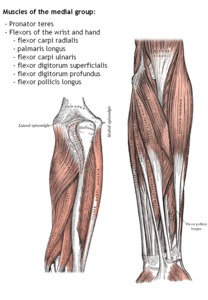
Fig. 6: Posterior and anterior views of left elbow and forearm showing the muscles of the medial group - Adapted from Gray's Anatomy 20th edition (1918) with original labels
(some nomenclature may not be up to date)
References: Public Domain (PD)
2.4.
Posterior group
- Triceps - the main extensor of the forearm,
is a strong muscle composed by three bellies; the long head originates from the infraglenoid tubercle; the lateral head from the posterior and lateral surface of humerus and the lateral intermuscular septum; the medial head has it's origin on the posterior surface of the humerus and lateral intermuscular septum,
both medial to the lateral head.
These three bellies coalesce distally to form the tendon of the triceps which attach to the olecranon and antebrachial fascia.
- Anconeus - a small triangular muscle with origin on the posterior surface of the lateral epicondyle,
travels distally to insert on the lateral posterior surface of the olecranon.
(also represented in Fig.
5)
3.
Main vessels and nerves
3.1.
Arteries
- Brachial artery - is the main artery present about the elbow.
It enters the cubital fossa descending from the upper arm,
marginating the medial border of the biceps brachii and the brachialis muscles,
and positions at middle of the cubital fossa,
passing between the bicipital aponeurosis and tendon.
- Radial artery - generally originates from the brachial artery 1 to 2 cm after passing the elbow joint.
It descends the forearm between the brachioradialis and the pronator teres muscles.
- Ulnar artery - deep in the cubital fossa,
the brachial artery stem divides and gives rise to the ulnar artery and common interosseous arteries.
Then the ulnar artery travels distally,
deeply to the pronator teres,
FCR,
PL and FDS muscles.
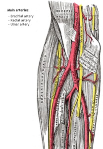
Fig. 7: Anterior view of left elbow. The main arteries about the elbow are represented - Adapted from Gray's Anatomy 20th edition (1918) with original labels
(some nomenclature may not be up to date)
References: Public Domain (PD)
3.2.
Veins
- Deep veins - these veins are considered vena comitants and accompany the described arteries,
generally in pairs (two veins to each artery),
they anastomose between each other and with superficial veins
- Superficial veins - veins traveling in the subcutaneous tissue,
the main superficial veins that cross the elbow are the basilic and cephalic veins,
basilic passes anteromedially and the cephalic anterolaterally.
On the majority of individuals,
a medial cubital vein connects these two,
at the elbow bend.
The network of superficial veins is highly variable between individuals.
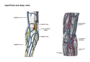
Fig. 8: Anterior views of the right and left elbow. The main superficial and deep veins are represented (respectively) - Adapted from Gray's Anatomy 20th edition (1918) with original labels
(some nomenclature may not be up to date)
References: Public Domain (PD)
3.3.
Nerves
(also represented in Fig.
7)
There are three major peripheral nerves in the elbow:
- Median nerve - follows the course of the brachial artery,
anterior to the artery at the cubital fossa,
passes between the two heads of the pronator teres muscle as it deepens.
- Ulnar nerve - located on the posteromedial aspect of the elbow,
in a sulcus composed by the olecranon and the medial epicondyle,
superficial to the posterior part of the ulnar collateral ligament.
Then,
follows distally between the two heads of the FCU.
- Radial nerve - courses between the brachialis and the brachioradialis,
passes deeply,
over the lateral epicondyle and divides into it’s superficial and deep branches after the elbow.

 with original labels
(some nomenclature may not be up to date) References: Public Domain (PD)")
 with original labels
(some nomenclature may not be up to date) References: Public Domain (PD)")
 with original labels
(some nomenclature may not be up to date) References: Public Domain (PD)")
 with original labels
(some nomenclature may not be up to date) References: Public Domain (PD)")
 with original labels
(some nomenclature may not be up to date) References: Public Domain (PD)")
 with original labels
(some nomenclature may not be up to date) References: Public Domain (PD)")
 with original labels
(some nomenclature may not be up to date) References: Public Domain (PD)")
 - Adapted from Gray's Anatomy 20th edition (1918) with original labels
(some nomenclature may not be up to date) References: Public Domain (PD)")