ECR 2016 / C-0400
The tale of global hypoxic ischaemic injury
This poster is published under an open license. Please read the disclaimer for further details.
Congress:
ECR 2016
Poster Number:
C-0400
Type:
Educational Exhibit
Keywords:
CNS, CT, MR, Education, Computer Applications-Detection, diagnosis, Imaging sequences, Acute, Ischaemia / Infarction
Authors:
L. M. Zammit1, R. Grech2; 1Paola/MT, 2Dublin 9/IE
DOI:
10.1594/ecr2016/C-0400

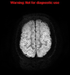
Fig. 1:
DWI MR, at 24 hours: Cortical laminar necrosis.

Fig. 2:
DWI MR, at 48 hours: Cortical laminar necrosis.
Gray matter and basal ganglia...

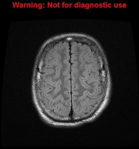
Fig. 3:
Flair MR, at 24 hours: Cortical swelling.

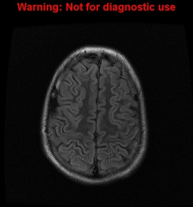
Fig. 4:
FLAIR MR, at 48 hours: Cortical swelling.

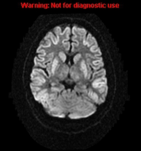
Fig. 5:
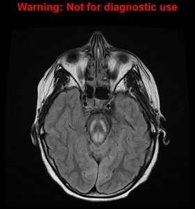
FLAIR , at 12 hours: Face-of-panda sign.

Fig. 6:
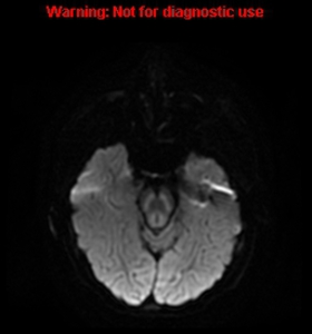
DWI MR, at 12 hours: Face of Panda sign.

Fig. 7:
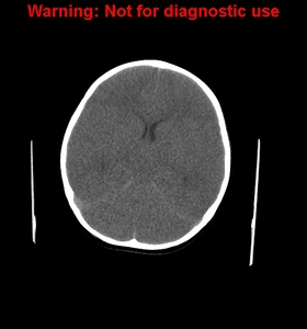
CT, at 6 hours: Global oedema with pseudo-subarachnoid sign.
 References: Medical Imaging Department, Mater Dei Hospital, Malta, EU")
Fig. 8:
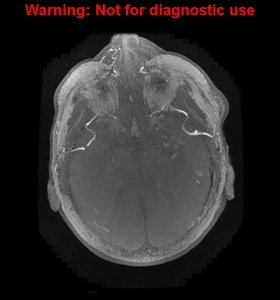
TOF MRA, at 6 hours: Absence of flow within ICAs but preserved in ECA : red...