This poster is published under an
open license. Please read the
disclaimer for further details.
Type:
Educational Exhibit
Keywords:
Fistula, Dissection, Aneurysms, Stents, Shunts, Embolisation, MR-Angiography, MR, Vascular, Neuroradiology brain, Interventional vascular
Authors:
M. Suzuki1, R. Irie2, N. Takano1, M. Hori2, K. Kamagata2, K. Kumamaru2, M. Yamamoto2, H. Ohishi2, S. Aoki2; 1Tokyo/JP, 2Tokyo, Japan/JP
DOI:
10.1594/ecr2016/C-0556
Background
TOF-MRA is,
- Based on the principle of flow related enhancement,
and since flow signal is sensitive to saturation effects,
slow flow can be difficult to clearly visualize.
- In addition,
faster,
or turbulent flow,
may cause a loss of signal or a vessel parallel to the scan-plane may result in signal loss from the vessel.
- Visualizing flow means visualizing arterial geometry and patency.
SILENT MRA is,
- Insensitive for saturation effects and it helps to visualize a vessel,
even when the flow is moving slowly,
regardless of its direction
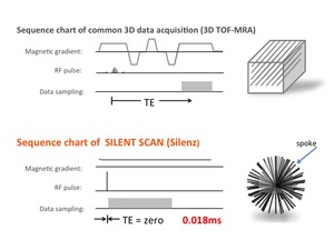
Fig. 1: pulse sequence charts of two MRAs
- SCAN detail,
- The blood within the carotid arteries is “tagged” using a long RF inversion pulse commonly referred to as a “Labeling ” pulse.
Once the blood is tagged,
it is allowed to flow into the vasculature and captured by the Silenz acquisition.
- This is followed by the collection of a control dataset where a “Labeling” pulse is applied above the head to minimize magnetization transfer effects and to control artifacts.
- These two datasets are subtracted to eliminate the background,
leaving a depiction of the entire vascular tree.
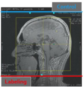
Fig. 2: indication of labeling slice and control slice
Based on the algorithm,
we applied the SILENT MRA for several cerevrocvascular disease.
