Learning objectives
- Toreview MRI technique and protocol in the assessment of pelvic floor dysfunction (PDF).
- To identify signs that are useful for the surgeon
- To provide the key points for aradiological template
- To provide the key points for a radiological template.
Background
PFDs are a common clinical problem with a significant impact on the quality of life,
especially in women.
Moreover,
25% of population is affected by PFDs.
The first approach is the anamnesis and the physical examination.
However,
since abnormalities of the three pelvic compartments are frequently associated,
a complete evaluation of all the pelvis is needed to define the treatment strategy.
Defeco-MRI is a valuable diagnostic tool to carry out a correct assessment about the functional and anatomical disorders of the pelvic floor.
Findings and procedure details
Defeco-MRI allows seeing the squeezing,
straining and defecation of the patients in a dynamic mode.
The protocol provides for the use of phased array surface coil and the supine position of patient.
The rectal filling is mandatory; we use 180-200 cc of sonographic gel.
The protocol must include axial,
sagittal and coronal TSE-T2.
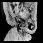
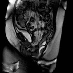
The steady-state sequence is used for the dynamic imaging,
acquiring one section per second in the midsagittal plane at rest,
during maximal sphincter contraction,
straining and defecation (figure 1).
Radiological template must...
Conclusion
PFDs are frequent and also complex conditions that can involve some or all pelvic viscera.
A structured report helps in this aim to give a complete evaluation which is needed to define the treatment strategy.
References
Koenraad J.
Mortele,
Janice Fairhurst.
Dynamic MR defecography of the posteriorcompartment: Indications,
techniques and MRI features.
European Journal of Radiology61 (2007) 462–472.
Alfonso Reginelli,
Graziella Di Grezia,Gianluca Gatta.,Role of conventional radiology and MRI defecography of pelvic floor hernias.
Reginelli et al.
BMC Surgery 2013,
13(Suppl 2):S53
Niccoló Faccioli,
Alessio Comai,
Paride Mainardi.
Defecography: a practical approach.
Diagn Interv Radiol 2010; 16:209–216
Wallden L.
Defecation block in cases of rectogenital pouch.
Acta Chir Scand1952 (suppl 165): 1-121.
Yang A,
Mostwin JL,
Rosenshein NB,
Zerhouni EA.
Pelvic...


