ECR 2016 / C-1827
Symptomatic Accessory Ossicles: A Pictorial Review
This poster is published under an open license. Please read the disclaimer for further details.
Congress:
ECR 2016
Poster Number:
C-1827
Type:
Educational Exhibit
Keywords:
MR, CT, Conventional radiography, Musculoskeletal system, Musculoskeletal bone, Normal variants, Education and training
Authors:
J. Saraiva, C. Bilreiro, L. Silva, C. Carneiro, B. M. Q. Santos, M. O. E. Castro; Portimão/PT
DOI:
10.1594/ecr2016/C-1827

Fig. 2:
os trapezoideum secundarium

Fig. 3:
os trapezoideum secundarium

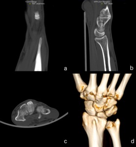
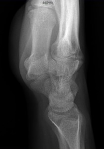
Fig. 4:
os styloideum

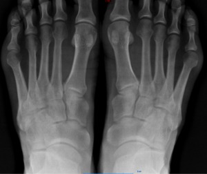
Fig. 5:
os intermetatarseum

Fig. 6:
os intermetatarseum

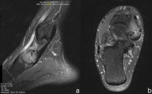
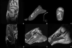
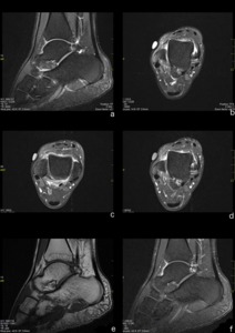
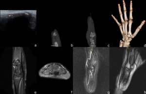
Fig. 7:
Accessory navicular

Fig. 8:
Accessory navicular

Fig. 9:
Accessory navicular

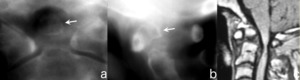
Fig. 10:
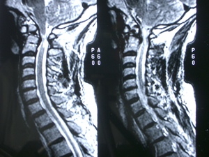
os odontoideum

Fig. 11:
os odontoideum

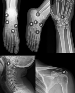
Fig. 1:
Locations of the more common symptomatic ossicles: 1. os odontoideum; 2....

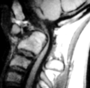
Fig. 12:
os odontoideum

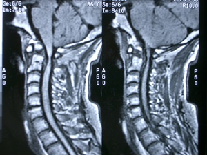
Fig. 13:
os odontoideum

Fig. 14

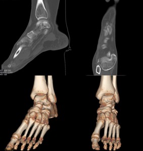
Fig. 15:
os trigonum

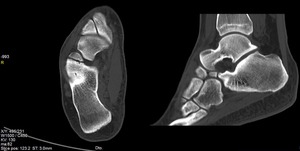
Fig. 16:
os calcaneus secundarius

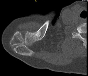
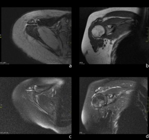
Fig. 17:
os acromiale

Fig. 18:
os acromiale