ECR 2017 / B-0772
Does the immunohistochemical pattern of breast cancer influence the detection by mammography, US or tomosynthesis?
This poster is published under an open license. Please read the disclaimer for further details.
Congress:
ECR 2017
Poster Number:
B-0772
Type:
Scientific Paper
Keywords:
Breast, Mammography, Ultrasound, Diagnostic procedure, Cancer, Molecular, genomics and proteomics
Authors:
P. Bartolomé, A. Quilez, F. Martinez Regueira, A. Fernandez Montero, A. Elizalde, L. J. Pina Insausti; Pamplona/ES
DOI:
10.1594/ecr2017/B-0772

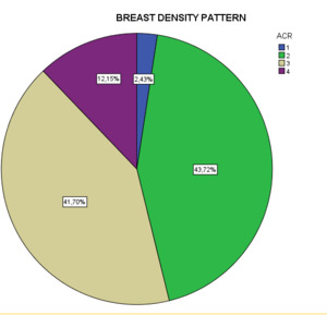
Fig. 1:
Distribution of breast density patterns in our sample

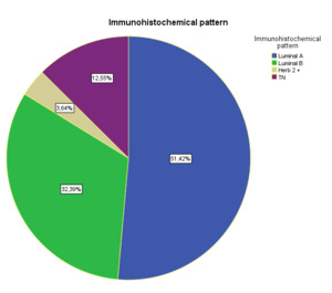
Fig. 2:
Distribution of immunohistochemical patterns of the tumors.

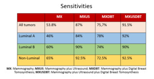
Fig. 3:
Summary of the sensitivities of the three techniques in our sample, according...

Fig. 5:
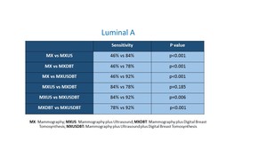
Performance of the different combinations of diagnostic techniques in Luminal A...

Fig. 6:
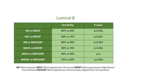
Performance of the different combinations of diagnostic techniques in Luminal B...

Fig. 7:
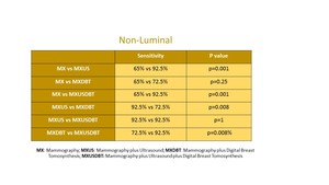
Performance of the different combinations of diagnostic techniques in...

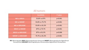
Fig. 4:
Performance of the different combinations of diagnostic techniques in all...

Fig. 9:
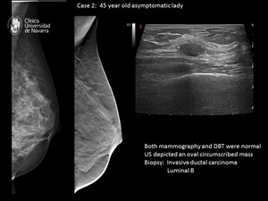
Case 2: Luminal B tumor only detected by US.

Fig. 10:
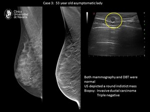
Case 2: Non-Luminal tumor only detected by US.

Fig. 8:
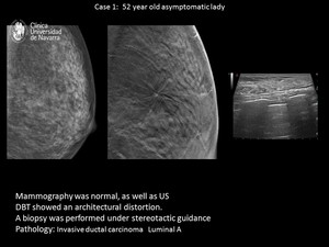
Case 1: Luminal A tumor only detected by DBT.