Learning objectives
To review the clinical indications,
technical considerations,
and pitfalls that should be considered in the acquisition,
processing,
and interpretation of the advanced MR images: magnetic resonance spectroscopy (MRS),
diffusion tensor imaging (DTI),
functional magnetic resonance imaging (fMRI),
perfusion-weighted imaging (PWI),
and voxel-based morphometry (VBM).
Background
Structural neuroimaging is highly important in the initial imaging approach of pathologies associated with the CNS because it allows to identify the localization and the anatomic relationship of brain lesions with other brain structures.
However,
advanced techniques are needed for a better characterization and classification.
Advances in neuroimaging during the last decades have improved the diagnostic and follow-up of patients.
The information obtained through these techniques goes beyond the anatomy and now it is possible to perform functional,
molecular,
and dynamic analysis of brain structures....
Findings and procedure details
MAGNETIC RESONANCE SPECTROSCOPY (MRS)
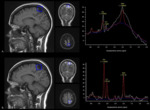
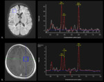
MRS is an imaging technique used to perform molecular analysis on brain tissue.
This MR modality allows to evaluate and to quantify the relative concentrations of different metabolites such as N-Acetylaspartate (NAA),
choline (Cho),
creatine (Cr),
myoinositol (mIns),
lipids,
and lactate.
MRS is a powerful tool for the assessment of metabolic disorders,
planning of image-guided biopsy,
and for the characterization,
classification,
and follow-up of brain lesions.
Moreover,
multiple studies support its use as abiomarker in the early and differential diagnosis in...
Conclusion
Knowledge of the clinical indications,
technical considerations,
and pitfalls of advanced neuroimaging is key in order to obtain high-quality images,
and to perform an accurate interpretation in the diagnostic approach of pathologies associated with the CNS.
Personal information
Ana María Granados Sánchez,
MD.
Neuro-radiologist
[email protected]
Juan Felipe Orejuela Zapata,
BSc.
Biomedical engineer
[email protected]
Sara Yukie Rodríguez Takeuchi,
MD.
Resident of radiology.
[email protected]
Radiology Department,
Fundación Valle del Lili.
Universidad ICESI.
References
AKSOY,
F.
G.,
LEV,
M.
H.
Dynamic contrast-enhanced brain perfusion imaging: technique and clinical applications.
Semin Ultrasound CT MR 2000; vol.21,
p.
462-477.
ALSOP,
D.
C.
Imagen de RM de perfusión.
En: Scott W,
ed Atlas.
Magnetic resonance imaging of the brain and spine.
3 ed.
Spanish edition.
Madrid: Marban; 2004.
p.
215-37.
Brandão L,
Domingues R.
MR spectroscopy of the brain.
1st ed.
Philadelphia: Lippincott Williams & Wilkins; 2004.
Cha S.
Perfusion MR imaging: basic principles and clinical applications.
Magnetic Resonance Imaging Clinics of...


