This poster is published under an
open license. Please read the
disclaimer for further details.
Type:
Educational Exhibit
Keywords:
Genital / Reproductive system female, Ultrasound, Diagnostic procedure, Congenital
Authors:
M. K. Vishweswaraiah1, P. G K2, P. Gowda2; 1Bangalore, Ka/IN, 2Bangalore/IN
DOI:
10.1594/ecr2017/C-1360
Findings and procedure details
The study was performed over a period of 6 months.
Initially conventional transabdominal and transvaginal ultrasonography was performed.
3-dimensional transvaginal utrasonography was performed using GE voluson 730 expert RIC5 9H transvaginal 4D probe on all cases suspected of MDA on transabdominal / transvaginal ultrasonography.
Volume was obtained using a sweep angle of 90° from one side of uterus to other.
Images were then reconstructed in coronal plane to the uterus using post processing technique.
Following were the spectrum of MDA included in our study
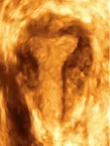
Fig. 6: Multiplanar reconstruction from 3-dimensional transvaginal ultrasonography with maximum intensity projection reconstructed image in a coronal plane to the uterus demonstrating normal uterus
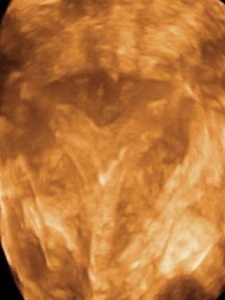
Fig. 7: Multiplanar reconstruction from 3-dimensional transvaginal ultrasonography with maximum intensity projection reconstructed image in a coronal plane to the uterus demonstrating Arcuate uterus
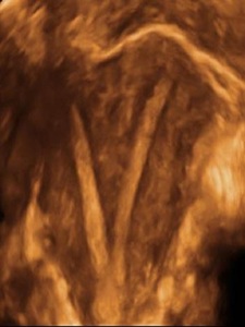
Fig. 8: Multiplanar reconstruction from 3-dimensional transvaginal ultrasonography with maximum intensity projection reconstructed image in a coronal plane to the uterus demonstrating bicornuate uterus
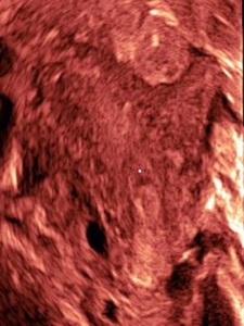
Fig. 9: Multiplanar reconstruction from 3-dimensional transvaginal ultrasonography with maximum intensity projection reconstructed image in a coronal plane to the uterus demonstrating unicornuate uterus with rudimentary horn

Fig. 11: Multiplanar reconstruction from 3-dimensional transvaginal ultrasonography with maximum intensity projection reconstructed image in a coronal plane to the uterus demonstrating septate uterus
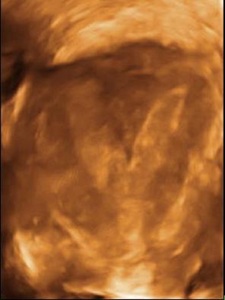
Fig. 12: Multiplanar reconstruction from 3-dimensional transvaginal ultrasonography with maximum intensity projection reconstructed image in a coronal plane to the uterus demonstrating subseptate uterus
