The technique: contrast agents and physical principles
Contrast-enhanced ultrasound (CEUS) involves the use of a microbubble contrast agent during an ultrasound exam.
These microbubbles contain low soluble gases encapsulated by a shell of phospholipids,
albumin or biopolymers.
They are designed to be smaller than 7 µm (the average size of a red blood cell) and exclusively intravascular.
The gas is eliminated through the airways 10-15 minutes after administration,
without renal or hepatic metabolism.
The key property of these microbubbles is their reaction when exposed to ultrasound.
Ultrasound beams with high mechanical index (MI) will lead to their destruction.
Ultrasound beams with low MI (0.08-0.2) will cause their nonlinear oscilation and consequent emission of harmonic echoes.

Fig. 1: Behaviour of microbubbles when exposed to ultrasounds. The nonlinear echo signal is due to the assymetrical fluctuation of diameter, with expansion being greater than compression.
Pulse Inversion technique (PI) allows separation of the harmonic echoes generated by the contrast within the blood stream from the echoes generated by the surrounding tissues.
In PI,
two ultrasound pulses are sent into the tissues,
being the second one a mirror image of the first.
These two pulses are then added at reception,
cancelling the linear signal from the surrounding tissues and exhibiting the non-linear signal from the microbubbles’ oscillation.

Fig. 2: Pulse Inversion technique. The sumation of two mirror pulses cancels the linear echoes from the tissues while still exhibiting the nonlinear echoes from the microbubbles.
The main advantages of CEUS over other techniques include its non-invasiveness,
safety and no exposure to radiation or nephrotoxic agents while delivering real time imaging of blood flow at a tissue perfusion level.
The major pitfalls are its dependence on the operator and poor performance when gas,
fat tissue or bone structures are scanned between the transducer and the region of interest.
Performing CEUS
The technical requirements needed to perform CEUS are an ultrasound equipment with contrast imaging capabilities,
microbubble contrast agent,
5 ml of saline solution and a patient with peripheral venous access and no contraindications for the administration of ultrasound contrast agents.
The understanding of CEUS’ physical principles and its structured examination protocol is essential to correctly perform the exam with reproducible results.
Our institution’s examination protocol is as follows:
1) Prepare the contrast,
according to the manufacturer instructions:
The mean recommended dose is 2,4 ml,
but it can be variable (1-4 ml),
allowing repetition of the injection if needed.
Check the contraindications given by the manufacturer,
mainly hypersensitivity to the active substance and severe cardiovascular disease (eg.
right-to-left shunts and severe pulmonary hypertension).
2) Establish the peripheral venous access:
Preferably the median cubital vein of the left arm,
for ergonomic purposes.
The minimum diameter of the catheter should be 20 gauge to avoid microbubble rupture.
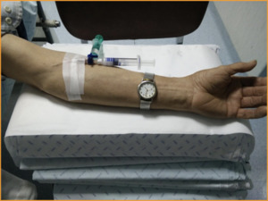
Fig. 3: Peripheral venous access (left arm) for CEUS examination.
3) Determine the region of interest to be studied:
Identify the area of study during B-mode exam.
Choose scanning planes that require the least collaboration from the patient (intercostal approaches are often helpful).
4) Start the exam
Set the ultrasound machine in contrast mode (low MI).
Inject the contrast followed by a 5 ml saline flush.
Start the timer immediately after injection.
Record the exam on video (in our experience,
it is common that definitive conclusions are only achieved after posterior reviewing the exam’s video).

 for CEUS examination.")

