ECR 2017 / C-2406
Increased nuchal translucency - Radiological and pathological review of a case series
This poster is published under an open license. Please read the disclaimer for further details.
Congress:
ECR 2017
Poster Number:
C-2406
Type:
Educational Exhibit
Keywords:
Obstetrics (Pregnancy / birth / postnatal period), Forensic / Necropsy studies, Foetal imaging, Ultrasound, Conventional radiography, Intrauterine diagnosis, Foetus, Pathology
Authors:
D. Castelo1, M. Marinho1, F. C. Pires1, S. Florim1, R. Nogueira2, C. Maciel3, F. Valente4; 1Vila Nova de Gaia/PT, 2Braga/PT, 3Porto/PT, 4Gaia/PT
DOI:
10.1594/ecr2017/C-2406
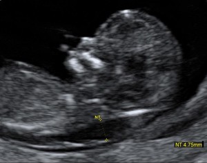
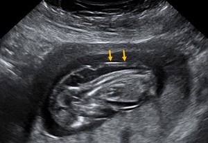
 proven to be a Down syndrome.")
Fig. 1:
Increased nuchal translucency at first trimester ultrasound (12 weeks) proven...
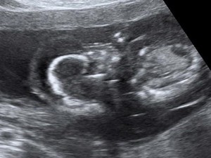
. Chorionic villus sampling revealed a Down syndrome.")
Fig. 5:
Mid-sagital plane at first trimester pregnancy. Note the cervical cystic...
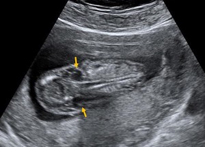
. First trimester ultrasound (12 weeks), axial plane at cranium base. Proven to be a case of Turner syndrome.")
Fig. 3:
Large cystic hygroma (arrows). First trimester ultrasound (12 weeks), axial...
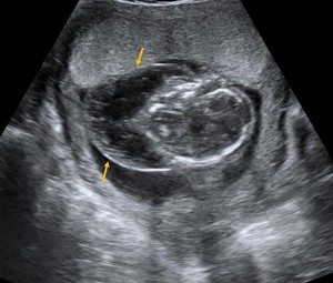
. Same case as above.")
Fig. 2:
Bilateral lateral neck cysts (arrows). Same case as above.
.")
Fig. 4:
Coronal plane of a Down syndrome case where a massive cystic hygroma involves...