ECR 2018 / C-0021
Epiploic appendagitis: CT imaging findings of an unusual cause of abdominal pain
Congress:
ECR 2018
Poster Number:
C-0021
Type:
Educational Exhibit
Keywords:
Inflammation, Acute, Contrast agent-intravenous, CT, Colon, Abdomen, Peritoneum
Authors:
D. Giambelluca1, S. Pellegrino1, G. Caruana1, M. Dimarco1, M. R. VACCARO NOTTE2, D. Picone1, M. Lenzo1, G. Lo Re1, R. Lagalla1; 1Palermo/IT, 2PALERMO, ITALIA/IT
DOI:
10.1594/ecr2018/C-0021
 that abuts colon-sigmoid junction.")
Fig. 1:
Primary epiploic appendagitis in 43-year-old man with clinical diagnosis of...
 and adjacent fat stranding (arrowhead).")
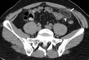
Fig. 2:
Acute epiploic appendagitis near the sigmoid colon in 41-year-old man. Axial...
 with fat density and surrounding inflammation (arrowhead) that abuts the hepatic flexure of transverse colon.")
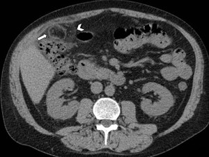
Fig. 3:
Acute epiploic appendagitis in a 53-year-old man. Axial non-contrast CT image...
 that abuts the transverse colon and has a central focal area of hyperattenuation (arrow) with surrounding fat inflammation (arrowhead).")
Fig. 4:
Acute epiploic appendagitis with a hyperattenuating center in a 40-year-old...
, that is depending directly from the vermiform appendix (curved arrow).")
Fig. 5:
Acute epiploic appendagitis near the vermiform appendix in 39-year-old woman....
 Axial CT scan of abdomen (bone window) shows a calcified, ring-like mass (arrow) medial to the sigmoid colon. It was initially mistaken for a mesenteric lymph node. B) Coronal reformatted CT image of the same patient shows the peritoneal loose body in the pelvic cavity (arrow).")
Fig. 6:
Calcified, amputated epiploic appendix in a 81-year-old man. A) Axial CT scan...