
ECR 2018 / C-1386
Abdominal Wall Endometriosis: MR Imaging Findings
Congress:
ECR 2018
Poster Number:
C-1386
Type:
Scientific Exhibit
Keywords:
Pathology, Diagnostic procedure, MR, Soft tissues / Skin, Pelvis, Musculoskeletal soft tissue
Authors:
B. S. Seker, G. YILMAZ, M. Atalar, B. Yıldız; SIVAS/TR
DOI:
10.1594/ecr2018/C-1386
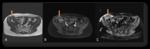
Fig. 3:
Case no: 7. A 24-year-old woman with a history of cesarean section. (A)...
 T2-weighted
coronal and (B) axial MR images show heterogeneous signal intensity with dark signal foci
at the anterior segment of left rectus abdominis muscle (white arrows). In addition, a
pfannenstiel incision scar is also seen (A, black arrow). (C) Post-contrast T1 weighted
image shows enhancement of this mass (white arrow) except for dark signal areas. (D)
ADC value is 1.09x10⁻³ mm2/s (white arrow)")
Fig. 4:
Case no: 11. A 33-year-old woman with a history of cesarean section. (A)...

Fig. 1
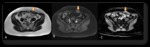
Fig. 2:
Case no: 4. A 29-year-old woman with a history of cesarean section. (A)...