ECR 2018 / C-1421
A retrospective study on surveillance of women with BRCA1 and BRCA2 mutation: outcomes from Veneto Institute of Oncology (IOV)
Congress:
ECR 2018
Poster Number:
C-1421
Type:
Scientific Exhibit
Keywords:
Breast, Mammography, MR, Ultrasound, Screening, Diagnostic procedure, Outcomes analysis, Cancer, Genetic defects
Authors:
A. Gaudino1, G. Romanucci2, E. Baldan1, S. Zovato1, F. Caumo1; 1Padua/IT, 2Verona/IT
DOI:
10.1594/ecr2018/C-1421

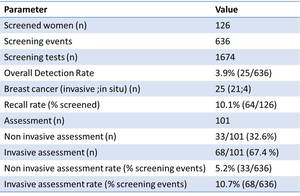
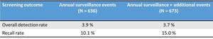
Table 1:
Main data and outcomes of the annual surveillance program of BRCA1/2 high-risk...

Table 2:
Shows the imaging modalities by which cancers were depicted.

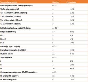
Table 3:
Characteristics of cancers found in the high-risk population.

Table 4

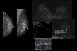
Fig. 1:
Example of true positive in a BRCA2 carrier with negative FFDM and suspicious...
 and suspicious MRI. The linear contrast enhancement was visible at the right side between the lower quadrants. A lesion with partially irregular margins was biopsied at the second-look US It was a benign lesion (focal sclerosing adenosis).")
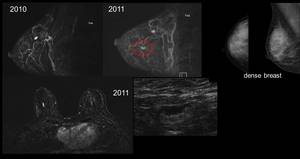
Fig. 2:
Example of false positive in a BRCA1 carrier with negative FFDM (dense breast)...