Type:
Educational Exhibit
Keywords:
Neoplasia, Cysts, Arteriovenous malformations, Education, Diagnostic procedure, MR, CT, Oncology, Eyes
Authors:
S. Hamid1, S. Arooj2, A. Waheed2, N. Ahmed2, T. Mahmood2; 1Karachi, Sindh/PK, 2Karachi/PK
DOI:
10.1594/ecr2018/C-1904
Background
Orbit is an anatomically complex region enclosing the globe,
extraocular muscles,
retro-orbital fat,
neurovascular structures,
lacrimal gland and connective tissues.
It is a small anatomical area with little free space and therefore mass lesions usually result in proptosis of the globe and can adversely affect visual acuity by causing pressure over the optic nerve or extraocular muscles.
Anatomy:
Contents of the orbit include: [Fig.
1,2]
- Globe with its contents
- Extraocular muscles (Superior,
Inferior,
Medial and Lateral Recti and Superior and inferior Oblique Muscles)
- Cranial nerves
- Autonomic nerves and ganglia
- Superior ophthalmic and infra-orbital arteries
- Superior and inferior ophthalmic vein
- Orbital fat
- Lacrimal gland
- Fascia bulbi (Tenon's capsule)
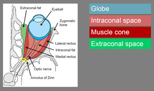
Fig. 1: Orbital Anatomy
References: Wichmann & Muller-Forell, Eur J Radiol. 2004
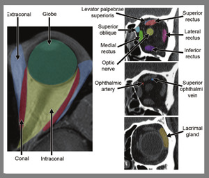
Fig. 2: Normal orbital anatomy. Axial CT image(left) with color overlays showing orbit divided into intraocular and extraocular spaces by the muscle cone and their relationship to the globe. Coronal CT images (right) with color overlays showing the configuration of the extraocular muscles, vascular structures, and lacrimal gland.
References: Tailor et al. (2013). Orbital Neoplasms in Adults: Clinical, Radiologic, and Pathologic Review. Radiographics : a review publication of the Radiological Society of North America, Inc. 33. 1739-1758. 10.1148/rg.336135502.
Orbital margins:
The bony margins of the orbit are composed of seven bones:
[Fig.
3]
- Pars orbitalis of the frontal bone
- Lacrimal bone
- Lamina papyracea of the ethmoid bone
- Orbital process of the zygomatic bone
- Orbital surface of the maxillary bone
- Orbital process of the palatine bone
- Greater and lesser wings of the sphenoid bone

Fig. 3: Bony Orbit
References: Radiology, University Institute for Radiodiagnostics Salzburg, Universitätsklinikum Salzburg - Salzburg/AT
Radiological Imaging:
Cross-sectional imaging is frequently utilized to help confirm the presence of an orbital mass and define its extent.
Characteristic imaging features of various patholgies help distinguish the lesions twith overlapping clinical presentations.
Both CT scan and MRI are frequently being used fo this porpose.
CT scan is most versatile in detecting bony details and calcification.
It has better temporal / spatial resolution.
However,
beam hardening artifacts from dental fillings often deteriorate the image.
MRI has inherent excellent soft tissue contrast and is best for evaluation of orbital soft tissues.
Especially the fat-saturation techniques improves the ability to visualize the optic nerve by suppressing the retro-orbital fat.
MRI is free from ionizing radiations.

