ECR 2018 / C-2007
Solitary Round Pulmonary Lesions in the Pediatric Population
Congress:
ECR 2018
Poster Number:
C-2007
Type:
Educational Exhibit
Keywords:
Paediatric, Respiratory system, Thorax, Conventional radiography, CT, MR, Diagnostic procedure, Laboratory tests, Education, Inflammation, Neoplasia, Congenital
Authors:
N. A. Arkoudis, A. Pastroma, G. Velonakis, V. Bizimi, C. Kontopoulou, K. Priftis, N. L. Kelekis, E. Alexopoulou; Athens/GR
DOI:
10.1594/ecr2018/C-2007
 in the pediatric population, as well as helpful clues for diagnosis. References: 2nd Department of Radiology, University General Hospital of Athens "Attikon"")
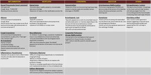
Table 2:
Conclusive table - take home messages - showcasing the differential diagnosis...