Type:
Educational Exhibit
Keywords:
Education and training, Technical aspects, Ultrasound, Musculoskeletal system, Musculoskeletal joint, Anatomy
Authors:
N. Pereira da Silva, J. A. Torres de Abreu Macedo, F. M. G. S. Pereira da Silva, F. Cruz, F. Caseiro Alves; Coimbra/PT
DOI:
10.1594/ecr2018/C-2114
Background
Ultrasound (US) has unique characteristics that make it very useful in the assessment of the musculoskeletal system,
namely good spatial resolution,
allowing immediate clinical correlation and dynamic assessment.
A more widespread access to high resolution ultrasound probes has raised interest in its application to smaller structures such as those present in the wrist.
In order to maximize the accuracy of wrist US,
radiologists must be comfortable with the complex anatomy of this joint,
with the more common clinical complaints and with the adequate examination technique.
Frequently,
wrist examination is directed toward a specific clinical question.
Despite this,
a more comprehensive exam may be considered.
Therefore,
the technique will be described based on a more thorough assessment of dorsal and ventral areas and of the comprising structures (Table.
1).
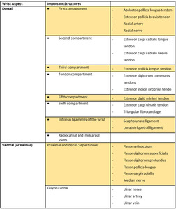
Table 1: Structures assessed in US of the wrist
