ECR 2018 / C-2167
Prostate gland multiparametric resonance. From guidelines to practice.
Congress:
ECR 2018
Poster Number:
C-2167
Type:
Educational Exhibit
Keywords:
MR, Oncology, MR-Functional imaging, Diagnostic procedure, Education, Cancer, Education and training
Authors:
C. Gómez 1, L. A. Ruiz Elizondo2; 1México /MX, 2Mexico City/MX
DOI:
10.1594/ecr2018/C-2167

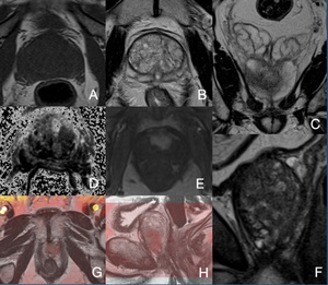
Fig. 6:
Prostate gland base. A. DWI B. ADC.

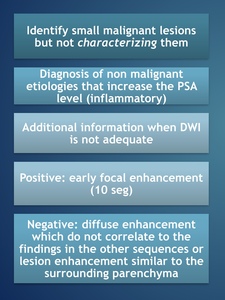
Fig. 7:
DCE 10 sec post-gadolinium administration

Table 1:
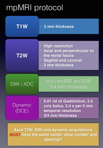
mpMRI indications, patient preparation and needed information.

Fig. 8:
T1WI.

Fig. 5:
Axial TW2 prostatic base.

Fig. 4:
Axial T1WI mid level.