Type:
Educational Exhibit
Keywords:
Abdomen, Anatomy, CT, Conventional radiography, Ultrasound, Education, Education and training, Peritoneum
Authors:
A. R. Ventosa, C. Carneiro, R. Monteiro, P. M. G. Alves, J. Brito; Algarve/PT
DOI:
10.1594/ecr2018/C-2542
Background
The falciform ligament is a sickle-shaped peritoneal double fold that attaches the liver to the anterior abdominal wall and the diaphragm.
It contains the ligamentum teres and the paraumbilical veins,
along with a variable amount of adipose tissue.
It is also the anatomical landmark responsible for the division between left and right subphrenic spaces.
The falciform ligament has three edges:
- The antero-superior or diaphragmatic edge attaches to the inferior surface of the anterior part of the diaphragm and to the inner surface of the anterior abdominal wall above the umbilicus.
This border is continuous with the diaphragmatic peritoneum and the anterior parietal peritoneum[1].
- The postero-inferior or hepatic edge attaches to the superior and anterior surfaces of the liver and to the fissure for ligamentum teres,
being continuous with the hepatic visceral peritoneum.
- The free edge connects inferiorly the diaphragmatic and hepatic edges.
It contains the ligamentum teres or round ligament,
that corresponds to the partially obliterated umbilical vein of the fetus [2,3].
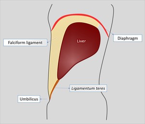
Fig. 1: Representative scheme of falciform ligament edges depicted in a sagittal plane. Note its sickle-shape.
References: Ana Rita Ventosa
Both right and left layers of the falciform ligament are continuous with the upper lamina of the coronary ligament[4].
Vascularization of the falciform ligament is highly variable.
The main arterial supply of the falciform ligament is provided by an artery that results from the anastomosis between the left inferior phrenic artery and a middle hepatic artery[1].
It has also been described the contribution of a terminal branch from the internal thoracic artery to its blood supply[5].
The venous drainage is provided by the paraumbilical veins,
a system of small veins around the falciform ligament,
which are divided in three groups[5,6]:
- Superior group of Sappey’s veins: this group drains the upper region of the falciform ligament and the medial portion of the diaphragm and enters the peripheral portal system of the left hepatic lobe.
This group communicates with branches of the superior epigastric or internal thoracic veins and with the inferior phrenic veins.
- Inferior group of Sappey’s veins: this group drains the inferior region of the falciform ligament,
also entering the peripheral portal system of the left hepatic lobe.
It communicates with branches of inferior epigastric veins.
- Burow’s veins – A pair of veins that communicates with branches of inferior epigastric veins and terminates in the middle portion of the collapsed umbilical vein,
not entering directly the portal system.
- Intercalary veins are small communicating branches between Burow’s and Sappey’s veins.
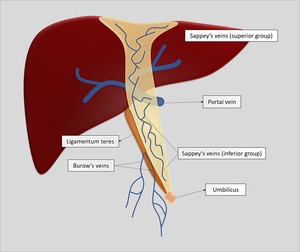
Fig. 2: Representative diagram of paraumbilical veins around the falciform ligament.
References: Ana Rita Ventosa
The falciform ligament may be involved by various pathologic conditions related to its peritoneal nature,
its content,
or by direct extension from neighboring pathologic processes.
Other disorders do not implicate the falciform ligament itself,
but either typically occur in its region,
such as some hepatic conditions,
or may result in peculiar imaging features,
such as air or fluid in subphrenic spaces.


