1. Rams horn stomach
Less than 4.00% patients of Crohn’s disease have involvement of the stomach and duodenum.it will cause conical narrowing,
tubular shape and limited distentibility of the stomach. The antrum is the gastric region most frequently involved.
These may give the stomach an unusual shape resembling the horn of a ram.
[1]
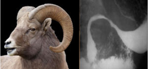
Fig. 1: Rams horn stomach
References: Baldwin M, Genant J, Braver J, Mortele KJ. Part 1-Classic signs in gastrointestinal radiology. Applied Radiology. 2011;40(12):22
2. Leather bottle stomach
A leather bottle appearance,
also known as linitis plastica.
The differential for the appearance of a leather bottle stomach include scirrhous metastases from lung,
colon,
and pancreatic carcinomas; lymphoma,
Crohn’s disease,
sarcoidosis,
and syphilis.
Typically,
the stomach is diffusely thickened with a small lumen.
[1]
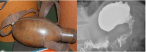
Fig. 2: Leather bottle stomach
References: https://radiopaedia.org/articles/linitis-plastica?lang=us
3. Windsock sign
it is a rare congenital cause of duodenal obstruction.
This windsock appearance is most commonly located in the second portion of the.
[1] it is a typical appearance of an intraluminal duodenal diverticulum on upper gastrointestinal contrast series which consists of an intraduodenal barium contrast-filled sac that is surrounded by a narrow lucent which is well demonstrated as the barium in the duodenum passes distal to the diverticulum.
[2]
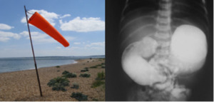
Fig. 3: Windsock sign
References: https://radiopaedia.org/articles/windsock-sign-duodenal-web
4. Double bubble sign
The double bubble sign represents the appearance of the distended gas and fluid-filled stomach and duodenum.
The double bubble sign indicates the presence of duodenal obstruction that can be caused by a duodenal web,
duodenal atresia,
and duodenal stenosis,
mal-rotation of the gut with a midgut volvulus or by Ladd bands,
or an annular pancreas.
[1]
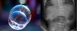
Fig. 4: Double bubble sign
References: https://radiopaedia.org/articles/double-bubble-sign-duodenum
5. Whirlpool sign
The whirlpool sign is found on CT and abdominal ultrasound in the presence of midgut volvulus,
caecal volvulus,
sigmoid volvulus,
closed loop bowel obstruction,
enteritis and omental torsion.
[2] it represents the swirling pattern of the gut and the superior mesenteric vein as they wrap around the superior mesenteric artery.
It also appears in ovarian torsion due to twisted vascular pedicle in colour doppler US study.
[1]
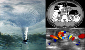
Fig. 5: Whirlpool sign
References: https://radiopaedia.org/articles/whirlpool-sign-mesentery?lang=us
6. String of pearls
This sign is also commonly referred to as the “string of beads” sign.
[1] It can be seen on upright or decubitus abdominal radiographs as well as on CT in patients with small bowel obstruction,
increased intraluminal fluid,
and slow resorption of intraluminal gas.
It consists of an obliquely or horizontally oriented row of small gas bubbles in the abdomen,
which represent small pockets of gas along the superior wall of the small bowel that are trapped between the valvulae conniventes.
The string of pearls sign,
when present in the appropriate clinical setting,
is virtually diagnostic of small bowel obstruction.
Although rare,
it may also be seen in adynamic ileus,
acute gastroenteritis,
and saline catharsis [2]
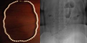
Fig. 6: String of pearls
References: https://radiopaedia.org/articles/string-of-pearls-sign-gastrointestinal
7. Stack of coins
The “stack of coins” sign typically indicates the presence of a small-bowel hematoma.
This sign is seen on plain films or MDCT images and represents adjacent,
thickened folds with sharp demarcation and crowding of the valvulae conniventes.
Other causes that may lead to the stack of coins sign include idiopathic thrombocytopenic purpura,
leukaemia,
pancreatitis,
pancreatic cancer,
haemophilia,
lymphoma,
myeloma,
chemotherapy,
and vasculidites.
[1]
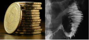
Fig. 7: Stack of coins
References: Baldwin M, Genant J, Braver J, Mortele KJ. Part 1-Classic signs in gastrointestinal radiology. Applied Radiology. 2011;40(12):22
8. String sign
In the setting of Crohn’s disease,
the terminal ileum often becomes markedly stenotic secondary to bowel-wall inflammation and fibrosis.
This results in the lumen of this portion of the small bowel resembling a piece of string on plain radiographs after ingestion of high-density oral contrast material.
it is also used for any severe narrowing of the bowel lumen,
including that seen in hypertrophic pyloric stenosis,
gastrointestinal tuberculosis,
carcinoid tumour and colon cancer.
[1]
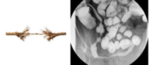
Fig. 8: String sign
References: https://radiopaedia.org/articles/string-sign-bowel?lang=us
9. Ribbon sign
Fluoroscopic examinations performed with high-density oral contrast material in patients with GVHD of the GI tract may demonstrate marked fold thickening,
luminal narrowing,
separation of folds,
and ultimately complete effacement of the valvulae conniventes.
The latter causes the so-called “ribbon sign”. The ribbon bowel appearance can also occur with multiple other clinical settings,
such as infection,
irradiation,
allergy,
ischemia,
ingestion of corrosives or medications,
amyloid,
mastocytosis,
lymphoma,
Crohn disease,
and celiac disease.
[1]
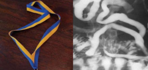
Fig. 9: Ribbon sign
References: Baldwin M, Genant J, Braver J, Mortele KJ. Part 1-Classic signs in gastrointestinal radiology. Applied Radiology. 2011;40(12):22
10. Comb sign
The comb sign refers to the hyper-vascular appearance of the mesentery in active Crohn disease.
This forms linear densities on the mesenteric side of the affected segments of small bowel,
which give the appearance of the teeth of a comb. The comb sign may be used to differentiate active inflammatory conditions from lymphoma and metastases,
which tend to be hypo-vascular.
[1]
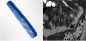
Fig. 10: Comb sign
References: https://radiopaedia.org/articles/comb-sign-mesentery
11. Bird’s beak sign
The “bird’s beak” sign is a classic finding on barium study; it describes a dilated proximal esophagus with a smooth-tapered,
distal esophagus at the level of the esophageal hiatus in the setting of achalasia.
The smooth tapering of the distal esophagus resembles the beak of a bird.
[1]
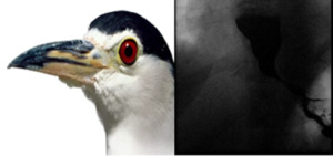
Fig. 11: Bird’s beak sign
References: https://radiopaedia.org/cases/achalasia-with-bird-beak-sign
12. Corkscrew sign
The “corkscrew” sign is the visual manifestation of lumen-obliterating,
simultaneous,
nonperistaltic contractions within the esophagus.These abnormal contractions of varying amplitude occur in diffuse esophageal spasm,
a rare esophageal motility disorder. Diffuse esophageal spasm is characterized on manometry by periods of normal peristalsis followed by simultaneous,
repetitive,
ineffective contractions.
These abnormal contractions segment the normal esophageal lumen,
mimicking a corkscrew on barium studies of the esophagus.
[1]
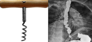
Fig. 12: Corkscrew sign
References: https://radiopaedia.org/cases/diffuse-oesophageal-spasm-corkscrew-oesophagus?lang=us
13. Bull’s eye lesions
Lesions within the stomach forming central collections of oral contrast within ulcerated intramural masses can produce a target or bull’s eye appearance on upper gastrointestinal barium examinations.
This differential diagnosis is includes gastric metastatic lesions from melanoma,
lymphoma, Kaposi’s sarcoma,
carcinoid tumours and gastric lipomas.
[1]
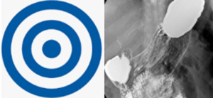
Fig. 13: Bull’s eye lesions
References: https://radiopaedia.org/cases/gastric-ulcer-a-bulls-eye-1?lang=us
14. Coiled spring sign
It is described as concentric ring shadows in a barium-filled cecum with a central filling defect.
These ring shadows represent contrast reflux within the lumen between the walls of the intussusceptum and intussuscipiens.
Classically,
this sign describes the appearance of the cecum in the presence of appendiceal intussusception,
a rare entity.
It is thought that the coiled-spring appearance results from intussusception of the cecal tip with the invaginated appendix acting as the lead point for variable amounts of cecocecal or cecocolic intussusception.
[3]
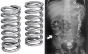
Fig. 14: Coiled spring sign
References: https://i.pinimg.com/474x/bd/26/a2/bd26a24fd3d2236a3fac20c4a54d5a04.jpg
15. Arrowhead sign
The arrowheadsign refers to the focal caecal thickening centred on the appendiceal orifice,
seen as a secondary sign in acute appendicitis.
The contrast material in the caecal lumen assumes an arrowhead configuration,
pointing at the appendix.
[3]
Fig. 15: Arrowhead sign
References: https://radiopaedia.org/cases/acute-appendicitis-with-ct-arrowhead-sign?lang=us
16. Thumbprint sign
Thumbprinting is a radiographic sign of large bowel wall thickening,
usually caused by edema,
related to an infective or inflammatory process (colitis).
Other condition like Pseudomembranous colitis,
ulcerative colitis,
lymphoma,
leukemia,
and hemorrhage into the bowel wall from coagulopathies may also produce this sign [3].
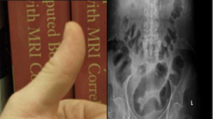
Fig. 16: Thumbprint sign
References: https://radiopaedia.org/articles/thumbprinting
17. Bowler hat sign
The “bowler-hat sign” represents the appearance of a sessile colonic polyp observed at an oblique angle on a double contrast barium enema.
The orientation of the dome of the bowler hat sign can help differentiate a polyp from a diverticulum.
Other intraluminal objects,
such as stool or air bubbles,
can also rarely create the bowler hat sign.
[3]
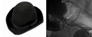
Fig. 17: Bowler hat sign
References: Miller Jr WT, Levine MS, Rubesin SE, Laufer I. Bowler-hat sign: a simple principle for differentiating polyps from diverticula. Radiology. 1989 Dec;173(3):615-7.
18. Accordion sign
The accordion sign is seen on CT examinations of the abdomen and refers to the similarity between the thickened edematous wall of pseudomembranous colitis and the folds of an accordion.
This appearance is the result of hyperaemic enhancing mucosa stretched over markedly thickened submucosal folds and is also seen when contrast is trapped between edematous haustral folds and pseudomembranes formed on the luminal surface of the colon.
[2]
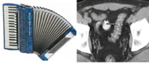
Fig. 18: Accordion sign
References: Yudin A. Metaphorical Signs in Computed Tomography of Chest and Abdomen. Springer International Publishing; 2014 Mar 10.
19. Lead pipe sign
The lead pipe appearance of colon is the classical barium enema finding in chronic ulcerative colitis.
There is complete loss of haustral markings in the diseased section of colon,
and the organ appears smooth-walled and cylindrical.
The differential diagnosis for a lead pipe appearance of the colon includes Crohn’s disease,
cathartic colon,
tuberculosis,
and,
rarely,
amebiasis.
[3]
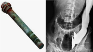
Fig. 19: Lead pipe sign
References: https://radiopaedia.org/articles/lead-pipe-sign-colon
20. Coffee bean sign
The coffee-bean sign is a sign on an abdominal plain radiograph of a sigmoid volvulus although some authors have also used the term to refer to closed loop small bowel obstructions. Caecal volvulus may be mistaken with sigmoid volvulus and the number of air-fluid levels help in differentiating the two entities.
[2] The coffee bean sign has also been used to describe small-bowel,
closed-loop obstructions.
[4]

Fig. 20: Coffee bean sign
References: https://radiopaedia.org/articles/lead-pipe-sign-colon
21. Mercedes-Benz sign
In the gallbladder,
the Mercedes-Benz sign describes a star-shaped pattern of gas-fissuring within gallstones initially described on an abdominal radiograph.Fissures,
usually fluid-filled,
are present in close to 50% of gallstones.
Less than half of these fissured gallstones contain some amount of gas.
The radiolucency caused by the gas usually appears in a triradiate pattern,
mimicking the Mercedes-Benz logo.
[2]
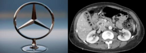
Fig. 21: Mercedes-Benz sign
References: https://radiopaedia.org/articles/mercedes-benz-sign-gallbladder
22. Champagne sign
The goblet sign (or champagne glass sign) refers to the appearance of the ureter when it is focally dilated by an intraluminal mass.
It is best seen when the ureter is opacified from below,
by a retrograde ureterogram.
Presence of this sign indicates the pathology to be chronic,
permitting the lesion to be accommodated in the ureter.
[2]
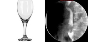
Fig. 22: Champagne sign
References: https://radiopaedia.org/articles/goblet-sign-ureter
23. Double duct sign
The double duct sign refers to the presence of simultaneous dilatation of the common bile and pancreatic ducts.
Being an anatomical sign it can be seen on all modalities that can visualize the region,
including: MRI,
CT,
ultrasound and endoscopic retrograde cholangiopancreatography (ERCP). The two most common causes of the double duct sign are carcinoma of the head of the pancreas and ampullary tumours (e.g. carcinoma of the ampulla of Vater),
although occasionally an impacted gall stone in the distal duct,
with associated edema,
can also result in obstruction of the pancreatic duct.[2]
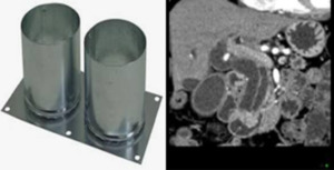
Fig. 23: Double duct sign
References: https://radiopaedia.org/articles/double-duct-sign?lang=us
24. Football sign
The football sign is seen in cases of massive pneumoperitoneum,
where the abdominal cavity is outlined by gas from a perforated viscus.
The median umbilical ligament and falciform ligament are sometimes included in the description of this sign,
as representing the sutures.[2]
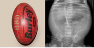
Fig. 24: Football sign
References: https://radiopaedia.org/articles/football-sign-pneumoperitoneum?lang=us

:22")


