ECR 2019 / C-1212
Imaging features of pancreatic neuroendocrine tumors: a 14 series case.
Congress:
ECR 2019
Poster Number:
C-1212
Type:
Educational Exhibit
Keywords:
Neoplasia, Endocrine disorders, Cancer, Contrast agent-intravenous, Ultrasound, MR, CT, Pancreas, Oncology, Abdomen
Authors:
J. Sanchez Dalmau1, M. Babarro Peleteiro1, P. Gómez Iglesias2, M. L. Rodriguez Rodil2; 1Madrid/ES, 2Móstoles/ES
DOI:
10.26044/ecr2019/C-1212

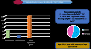
Fig. 13:
Types of pNETs and epidemiology in our study

Fig. 14:
Past medical history and symptomatology of presentation

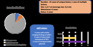
Fig. 15:
Imaging findings and pancreatic mimics of pNET in our institution.