Type:
Educational Exhibit
Keywords:
Infection, Cancer, Diagnostic procedure, Decision analysis, Computer Applications-Detection, diagnosis, CT, Conventional radiography, Thorax, Respiratory system
Authors:
N. Colic1, R. STEVIC2; 1Beograd, Serbia/RS, 2Belgrade/RS
DOI:
10.26044/ecr2019/C-1356
Background
Nodular changes are multiple round opacities,
in most cases of a diameter of 0.5 to 5mm
Precisely diagnosed with CT scan (superposition on X ray)
Nodular changes can be:
Milliar – fom 0.1-0.5mm lat.-millium
Micronodular- from 0.5 to 2 mm,
the size of the grain of the spring,
Macronodular - 2 to 5 mm
There are also round changes (5mm to 3cm) and tumor shadows (over 3cm)
Nodules are further characterized by their edges (clear or unclear),
the intensity of the shadow (semitransparent,
mycotic,
spot) and the distribution in the lungs (x ray or CT distribution).
Nodular changes are a radiological picture of pathological changes in acinus in numerous diseases of alveolar and interstitial origin.
During the pathological process in acinus there is the accumulation of transudate,
exudate,
hemorrhage,
cellular infiltration or there is accumulation of aspiration of different contents.
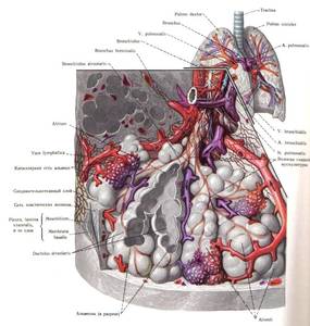
Fig. 1
