ECR 2019 / C-1718
Diffuse peritoneal disease: There is life beyond peritoneal carcinomatosis.
Congress:
ECR 2019
Poster Number:
C-1718
Type:
Educational Exhibit
Keywords:
Peritoneum, Genital / Reproductive system female, Gastrointestinal tract, CT, MR, Computer Applications-Detection, diagnosis, Education, Diagnostic procedure, Abscess, Multidisciplinary cancer care, Inflammation
Authors:
S. Belião1, J. Ip2, A. Gaivao2; 1Lisboa/PT, 2Lisbon/PT
DOI:
10.26044/ecr2019/C-1718

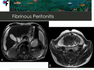
Fig. 2:
68-year-old patient with fibrinous peritonitis. Abdominal and Pelvic MRI T2WI...

Fig. 3:
68-year-old patient with fibrinous peritonitis. Abdominal MRI T1WI with fat...
. Focal thicker segments show obtuse angle with the peritoneum. References: Department of Radiology, Champalimaud Foundation, Lisbon")
Fig. 4:
Nonuniform peritoneal thickening in a 56-year-old female patient with...
 and absent peritoneal thickening.
References: Department of Radiology, Champalimaud Foundation, Lisbon")
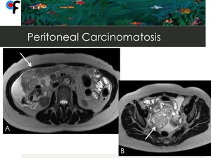
Fig. 5:
63-year-old female patient with ovarian cancer and peritoneal carcinomatosis....
 and a complex multiloculated cystic lesion at the right ovary (B). References: Department of Radiology, Champalimaud Foundation, Lisbon")
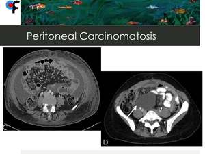
Fig. 6:
63-year-old female patient with ovarian cancer. Abdominal and pelvic MRI show...

Fig. 7:
A- Involvement of small bowel mesentery in a 70-year-old female patient with...

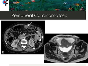
Fig. 8:
E- Presence of implant located at the anterior parietal peritoneum in a female...