This case series includes 10 patients with head and neck paragangliomas,
out of which 4 underwent MRI and 6 underwent CT.
MRI protocol included T1,
T2,
FLAIR,
DWI,
SWI and post contrast T1 weighted sequences.
CT protocol included thin section spiral CT (single slice and multidetector 128 slice) of the head and neck region with post contrast image acquisition for all cases expect 2 cases which were plain studies.
High resolution CT images were acquired for 2 patients.
On MRI,
paragangliomas showed variable signal on T1 and T2 sequences and high signal on FLAIR sequence with enhancement on post-contrast images and clear delineation of soft tissue extension as compared to CT.
Hypodense lesions,
heterogenously enhancing lesions,
lytic destruction of underlying bones,
widening of the jugular foramen,
erosion of carotid and facial canals were some of the imaging features on CT.
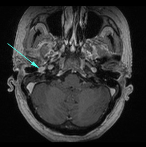
Fig. 1: T1 post contrast image showing intensely enhancing lobulated lesion in the right middle ear cavity adjacent to the tympanic membrane - glomus tympanicum
References: Radiodiagnosis & Imaging, M. S. Ramaiah Medical College and Hospitals, M. S. Ramaiah Medical Hospital - Bengaluru/IN
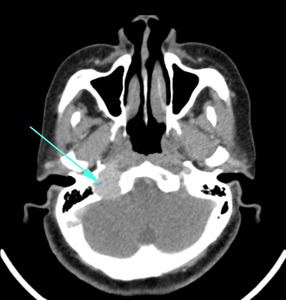
Fig. 2: Post contrast CT image showing an enhancing solid lesion in the region of the right jugular bulb eroding the petrous part of the right temporal bone and widening the jugular foramen - Case of glomus jugulare.
References: Radiodiagnosis & Imaging, M. S. Ramaiah Medical College and Hospitals, M. S. Ramaiah Medical Hospital - Bengaluru/IN
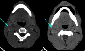
Fig. 3: Post contrast CT images showing heterogeneously enhancing lesion on the right side of the neck splaying the carotid bifurcation, abutting the sternocleidomastoid muscle laterally and trapezius medially - A case of carotid body tumour.
References: Radiodiagnosis & Imaging, M. S. Ramaiah Medical College and Hospitals, M. S. Ramaiah Medical Hospital - Bengaluru/IN
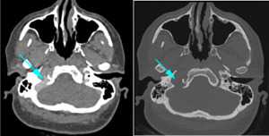
Fig. 4: Case of glomus jugulare: Contrast enhanced CT image showing enhancing lesion in the right internal jugular vein causing widening of the jugular foramen.
Bone CT axial image showing widening of the jugular foramen with erosion of the jugular spine.
References: Radiodiagnosis & Imaging, M. S. Ramaiah Medical College and Hospitals, M. S. Ramaiah Medical Hospital - Bengaluru/IN
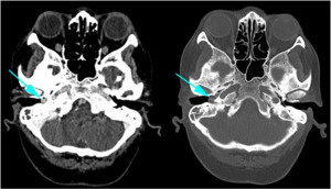
Fig. 5: Case of glomus tympanicum - Axial CECT and bone CT images showing enhancing soft tissue lesion in the right tympanic cavity causing erosions of the lateral wall of right carotid canal and facial canal.
References: Radiodiagnosis & Imaging, M. S. Ramaiah Medical College and Hospitals, M. S. Ramaiah Medical Hospital - Bengaluru/IN
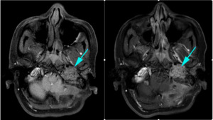
Fig. 6: Pre and post contrast T1 weighted images showing irregular hypointense enhancing lesion in the left jugular fossa - Glomus jugulare.
References: Radiodiagnosis & Imaging, M. S. Ramaiah Medical College and Hospitals, M. S. Ramaiah Medical Hospital - Bengaluru/IN
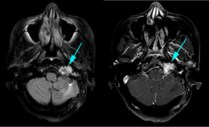
Fig. 7: Axial FLAIR and post contrast T1 weighted image showing residual / recurrent lesion in the left jugular fossa in a patient with known left glomus jugulare post excision.
References: Radiodiagnosis & Imaging, M. S. Ramaiah Medical College and Hospitals, M. S. Ramaiah Medical Hospital - Bengaluru/IN
