54 patients aged 25 to 57 years who underwent MRI examination of the brain using non-contrast angiography in our clinic in the period from 17.05.18 to 29.09.2018 who were included in the study.
Patients were without a history of atherosclerosis of the carotid in anamnesis have been included in the study.
The study was performed using 1.5 Tesla MR-scanner according to the standard protocol used in our clinic for MRI angiography of the cerebral arteries.
We used 3-dimensional (3D) time-of-flight (TOF) impulse sequence with 1 mm slice thickness,
256x228 mm matrix,
25 g angle,
repetition time/echo time 20/2.7 ms.
The scan region included the posterior cranial fossa,
skull base and COW area (40 mm above the skull base).
The absence of Pcomm visualization was not considered as aplasia based on the last publications data about the low specificity of MRI angiography in their determining [4].
The Pcomm which were differenciating on 2D images were analyzed by maximal diameter which was recorded [figure 1].
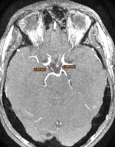
Fig. 1: Left and right Pcomm`s on axial M3D/TOF /SP/GP are detected in maximal diameter 1,5 mm and 1,8 mm.
References: Department of Radiology, MEDSI, Botkin hospital, Moscow
According to the results of measurements Pcomm`s were classified into 3 groups by visualization types [Table 1]:
Table 1. Pcomm visibility classification
|
Group
|
Pcomm blood flow MR-signal
|
Pcomm diameter
|
|
1
|
Not defined
|
Unknown
|
|
2
|
Traceable on 2D
|
<1,5 mm
|
|
3
|
Traceable on 2D and 3D
|
>1,5 mm
|
Besides Pcomm`s in each patient were explored cerebral arteries are illustrated on the scheme [figure 1]:
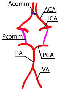
Fig. 2: Scheme of cerebral arteries which were explored in the study.
References: Department of Radiology, MEDSI, Botkin hospital, Moscow
· Anterior communicating artery (Acomm)
· Posterior communicating arteries (Pcomm)
· Ophthalmic segments of internal carotid artery (ICA)
· Anterior cerebral arteries (ACA)
· Posterior cerebral arteries (PCA)
· Basilar artery (BA)
· Vertebral arteries (VA)
Paired arteries of COW and intracranial segments of the vertebral arteries were analyzed by diameter and visibility on MRA.
The arterial hypoplasia was considered if there was difference of the diameters and the MR-signal intensity of the cognominal arteries.
Correlation of abovementioned arteries anatomy and the Pcomm visibility was estimated in each case.
Basilar artery (BA) and Anterior communicating artery (Acomm) were not evaluated in our study as they are unpaired.
There were implied that they had normal structure.


