ECR 2019 / C-2209
MRCP implementation methods in our institution
Congress:
ECR 2019
Poster Number:
C-2209
Type:
Scientific Exhibit
Keywords:
Radiographers, Biliary Tract / Gallbladder, MR, MR-Cholangiography, Contrast agent-oral, Contrast agent-intravenous, Cholangiography, Obstruction / Occlusion
Authors:
B. Zólyomi, I. Kéki; Budapest/HU
DOI:
10.26044/ecr2019/C-2209

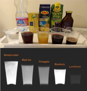
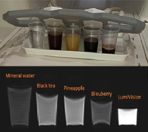
Fig. 3:
Representation of the whole biliary system without and after with LumiVision®

Fig. 5:
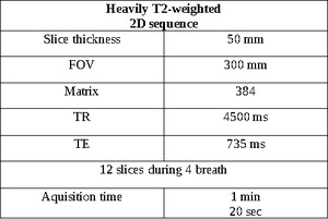
Radial planning of heavily T2-weighted 2D sequence

Fig. 6:
Imaging of the bile ducts with heavily T2-weighted 2D sequence
")
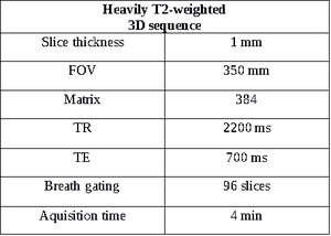
Fig. 8:
Imaging of the gallbladder and the biliary system with heavily T2-weighted 3D...
")
Fig. 9:
Imaging of the gallbladder and the biliary system with heavily T2-weighted 3D...

Fig. 11:
Raw image of T1-weighted thin sliced sequence in biliary phase

Fig. 12:
MIP image of T1-weighted sequence in biliary phase

Fig. 14:
High signal intensity of the hepato-specific contrast agent in the bile ducts,...