ECR 2019 / C-2630
Differentiation between clear cell renal cell carcinoma from other renal cell carcinoma subtypes: qualitative and quantitative multiphasic CT analysis
Congress:
ECR 2019
Poster Number:
C-2630
Type:
Scientific Exhibit
Keywords:
Pathology, Neoplasia, Efficacy studies, CT, Urinary Tract / Bladder, Kidney, Abdomen
Authors:
M. H. Verussa1, F. M. A. Coelho2, M. FUKUMOTO1, B. A. Vento1, R. Gianordoli1, S. Horta1, F. I. Yamauchi1, P. Viana1; 1São Paulo/BR, 2São Paulo, São Paulo/BR
DOI:
10.26044/ecr2019/C-2630

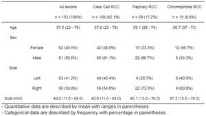
Table 1:
Epidemiology characteristics of all lesions and subgroups.

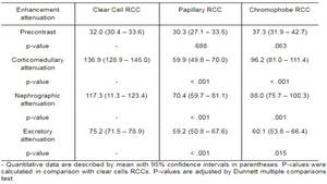
Table 2:
Attenuation of renal masses on the basis of histologic subtype.

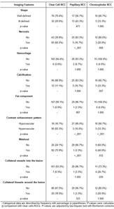
Table 3:
Imaging features of renal masses on the basis of histologic subtype.