Study population:
Prospective study of 74 Antenatal mothers[26 of them are primi and 48 of them are multiparous](Fig.1) without riskfactors with ultrasonically labelled normal fetuses of gestational age of 22 – 36weeks(Fig.2), Antenatal mothers with fetuses showing normal findings on sonographic brain examinations who gives complete informed consent to do foetal MRI ,
Familial history of gyrational abnormalities,
history of epilepsy,
Obese mothers in whom ultrasound not useful in assessing foetus,
mothers with epilepsy,
family history of epilepsy .
Antenatal mothers with twin gestation ,pregnancy induced hypertension ,gestational diabetes mellitus ,
intrauterine growth restriction are excluded from study .Gestational age distribution
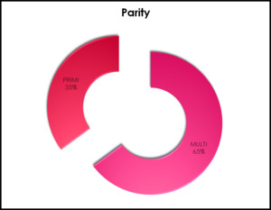
Fig. 1: Study population
Data aquisition:
Foetal MRI is performed by 1.5 T superconducting magnet using a phased array torso surface coil.Basic sequence used was half fourier acquired single shot turbo spin echo [ HASTE ] which is a type T2 spin echo sequence [single shot fast spin echo sequence -SSFSE ] ,
so that to reduce the movement artifacts created by foetal movements and to improve image quality.
Initially localizer is obtained with large field of view ranging from 320–400 mm to assess the foetal position in three orthogonal planes .
Slice thickness was kept as 4 – 6 mm ,Matrix size 169x256 ,Field of view of 330-360 ,Flip angle 90 with acquisition time of 1 slice per sec .About 9to 19 slice of images were obtained with interslice thickness of less than 2 mm .
Data analysis:
All the cerebral sulci [17 sulci] were looked for in an orderly manner starting from sulci that present in medial cerebral surface,followed by ventral cerebral surface and then those present in vertex .
The identified sulci were categorized in to three catagories
- Present
- Absent
- Partially developed
Sulcus which may seen as a very shallow indentation without a clear CSF space in between are termed as partially developed .Sulcus which is well conspicuous with a clear CSF space in between were termed as present.
Statistical analysis:
Sequential appearnce of various sulci with respect to gestational age are represented in graphical distribution.
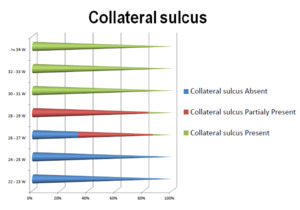
Fig. 3: Appearance of collateral sulcus with respect to gestational age
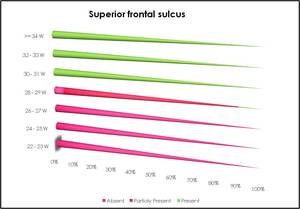
Fig. 4: Appearance of superior frontal sulcus with respect to gestational age
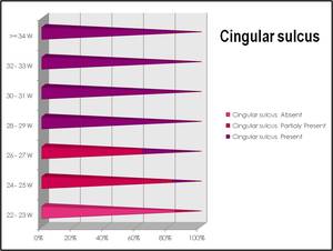
Fig. 5: Appearance of cingular sulcus with respect to gestational age
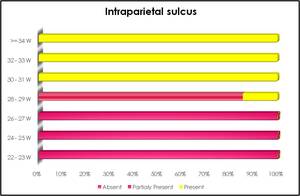
Fig. 6: Appearance of intraparietal sulcus with respect to gestational age


