Radiation protection survey and quality assurance of X-ray installations
The radiation exposure levels during the operation of the radiological equipment were measured for the radiological installation and quality control and quality assurance protocols prescribed by Atomic Energy Regulatory Board for x-ray equipment was carried out by using PTW NOMAX multimeter to ensure the stability,
filtration and output consistency.
The machine setup,
procedure,
and exposure protocols were optimized to be within tolerance limits as per AERB guidelines and a specialized radiation awareness programme was conducted for all radiation workers of the institution prior to the commencement of work.
Equipment
Alleger’s digital radiography (DR) machine (2015,
DIGIX,
India) with a 3-phase x-ray generator was used.
The measured Half Value Layer (HVL) for this machine is 3.0 mm Aluminium (Al) at 80 kV.
The tube outputs are between 40kVp and 150kVp with mAs range between .5 mAs and 500 mAs.
Dosimetry Apparatus
The use of Dose Area Product3 (DAP)meter is recommended as the most practicable to derive the Entrance Skin Dose (ESD) in radiological examination by many international agencies i.e IAEA/EU/AAPM and National agencies i.e AERB in their guideline on Diagnostic Reference Levels (DRL).
The major advantages of DAP compared to the other dosimetry tools are that it takes into account the whole area of examination irrespective of patient position in the beam,
the focus to film distance (FFD) and the non-interference in the radiographic examination of the patient.
In this study duly calibrated KermaX plus DAP meter of IBA Dosimetry,
Germany was used to measuring the absorbed dose in the air to arrive ESD value for Pediatric chest x-ray examination.
The DAP is placed in the frame of the collimator of x-ray machine and reading were obtained to calculate the entrance surface dose (ESD) for each chest examination as well as serves the input values of entrance air karma for calculation of gonadal dose through Monte Carlo simulation.
The Optically stimulated luminescence (OSL) dosimeter is another tool used for infield and out of field radiation dose measurement.
it is having good homogeneity with a lower systematic change in sensitivity from measurement to measurement of radiation up to certain range4.
To measure gonadal dose in a male child who underwent for chest radiography we used the Optically Simulated Luminescence Dosimeter (OSLD) Nanodots (Landauer,
2015) having a disc of 4mm diameter and 0.2 mm thickness mounted into the light-tight case.
The OSLD Nanodots are calibrated for use in diagnostic radiology energy range and dose value are obtained on Microstar OSL Nanodot reader of Landuar Inc.
For measurement of gonadal dose,
the nanodots were placed at the level of distal end of pubic symphysis joint of a male child for Anterior-posterior (AP) view of Chest and the level of Coccyx end of a male child for Posterior-anterior view of Chest in order to obtain the perpendicular orientation of OSLD disc for measurement.
the dose obtained from OSLD measurement were compared with calculated radiation dose value obtained from Monte Carlo simulation.
The Monte Carlo techniques based on calculations is another common method for evaluating radiation dose nowadays.
The Monte Carlo based software5PCXMC‑2 developed by STUK (Radiation and Nuclear Safety Authority in Finland),
was used to simulate projections and calculate the resulting effective doses from the projections.
The software calculated both organ doses for a large number of organs/tissues and the resulting effective dose to the patient using anatomical data from the mathematical phantom models.
This software uses organ weighting factors of both ICRP publication 60 and ICRP publication 103 for measurement of gonadal doses.
The calculations of gonadal dose in chest x-ray examination were carried out using the PCXMC-2 Monte Carlo radiation transport code on Intel ® Core™ i3 of 2.66 GHz CPU powered by HP having 8 GB RAM as installed memory.
To calculate Input data like SSD,
field size,
kVp,
Dose Area product mGy.cm2,
coordinates of the point inside the phantom through which point the central axis of the X‑ray beam is directed,
total filtration,
and anode angle are obtained at the time of study for each projection.
To mimic the real patient scaling of the inbuilt phantom was done by using child height and weight in six different phantom sizes,
representing patients of different ages,
from newborn to standard adult.
(Fig1)
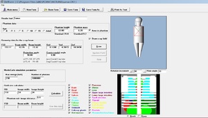
Fig. 1: PCXMC2.0 User Interface
References: PCXMC2.0 User Interface
Selection and Simulation of Patient
Random sample selection of pediatric male child was done and categorized into neonates (0-1yrs),
children (1-5years) and younger children ( 5-10 years) categories.
Routine chest X-ray examinations of pediatric patients were performed on Alleger’s digital radiography machine (2015,
DIGIX,
India) by placing OSLD Nanodots at level of distal end of pubic symphysis joint of male child for Anterior-posterior (AP) view of Chest and the level of Coccyx end of male child for Posterior-anterior view of Chest.
The study performed on 28 neonates 74 children and 10 younger children's,
and the chest x-ray exposure parameter was used in the range between 5mAs-32mAs & 52kVp-72kVp (Fig 2)
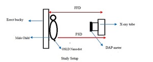
Fig. 2: Study Setup
Evaluation of gonadal dose and entrance surface dose (ESD) for Chest X-ray examination
The measurement of ESD calculation using DAP was conducted by using focus to film distance (FFD) is 180 cm.
The field size and focus to the skin (FSD) used in this study were measured during the experiment setup including the chest region of the male child.
DAP data was converted to the ESD (mGy) based on the equations suggested by Livingstone,
et al6.
(2006) and Meade,etal7(2003) The equation is as
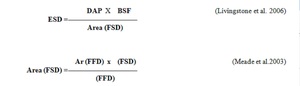
Fig. 3


