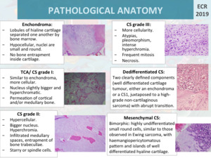
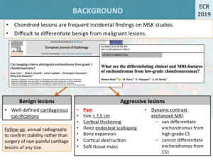
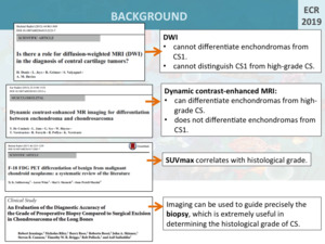
Chondroid lesions are frequent incidental findings on musculoskeletal studies. Both benign and malignant chondroid lesions are found most commonly in the diaphysis,
followed by the metaphysis of long bones.
A cartilage lesion in an elderly patient,
a painful injury and an injury in the axial skeleton are more likely to be malignant.
In fact,
the clinical finding of localized pain not attributable to other causes is the most useful indicator of chondrosarcoma [1].
Solitary lesions on the hands and feet are probably benign.
Chondroid matrix is usually not difficult to recognize,
but differentiating benign from malignant lesions is sometimes hardly difficult both radiographically and pathologically.
Fig. 4
However,
certain imaging findings can distinguish significantly between benign and aggressive lesions.
Longitudinal diameter greater than 7,5 cm,
cortical thickening,
deep endosteal scalloping,
bone expansion (except in fibula),
cortical breakthrough and soft tissue mass are characteristics that suggest malignancy.
On the other hand,
lesions with well-defined cartilaginous calcifications were less likely to be malignant [1,
2].
Plain radiographs,
CT and MRI are limited in their ability to distinguish benign from low-grade cartilage lesions,
although MRI has higher sensitivity than plain radiographs.
Fig. 2
Nevertheless,
MRI has limitations.
Diffusion weighted images (DWI) cannot differentiate between enchondromas,
grade 1 chondrosarcomas and high-grade chondrosarcomas [3].
Moreover,
in dynamic contrast-enhanced MR,
a pattern of peripheral enhancement around the cartilage lobes or a confluent enhancement pattern can differentiate enchondromas from high-grade chondrosarcomas,
but does not differentiate the enchondromas from grade 1 chondrosarcomas [1,
2,
4].
PET/CT is a useful technique together with conventional images in the characterization of chondroid tumours since the SUVmax correlates with the histological grade.
Very low SUVmax (<2.0) supports a diagnosis of benign tumour,
while elevated SUVmax (>4.4) is suggestive of higher-grade chondrosarcoma [5].
Furthermore,
imaging can be used to guide precisely the biopsy,
which is extremely useful in determining the histological grade of chondrosarcoma.
The accuracy of the diagnostic biopsy to distinguish low-grade from high-grade chondrosarcoma was 93.6% [6].
Fig. 3
Regarding follow-up,
annual radiographs of the cartilage lesion is recommended to confirm stability rather than surgery of non-painful cartilage lesions of any size [7].