Type:
Educational Exhibit
Keywords:
Genital / Reproductive system female, Oncology, MR, MR-Diffusion/Perfusion, Diagnostic procedure, Multidisciplinary cancer care
Authors:
H. Sahin, Y. Pekcevik; Izmir/TR
DOI:
10.26044/ecr2019/C-3664
Background
Borderline ovarian tumours (BOT) account for 15-20% of non-benign ovarian neoplasms [1].
These tumours show low malignant potential with intermediate mitotic activity,
nuclear atypia and lack of obvious invasion of the stroma [2].
They are most commonly serous or mucinous.
About 9-15% of all serous neoplasms are known to be borderline type [2](Figure 1).
Cystic serous tumours with more than 10% BOT architecture are classified as serous BOT according to last WHO classification of ovarian tumours (2014) [3].
Radiological diagnosis of serous borderline tumours is of paramount importance since they have a better clinical prognosis with high survival rates than their malignant counterparts,
even with intraperitoneal dissemination or lymph node metastasis.
SBOTs are usually seen in a younger age group,
typically between 40-50 years,
present at an early stage with a better prognosis when compared with malignant ovarian tumours [4].
Those factors make the way for fertility-sparing surgery as an important treatment option for patients with SBOT.
In this educational exhibit,
we aim to review MR imaging features and classification of SBOT according to imaging using our case series.
We aim to demonstrate malignant tumours mimicking SBOT and review the clinical approach to SBOT including staging,
management,
treatment and follow-up.
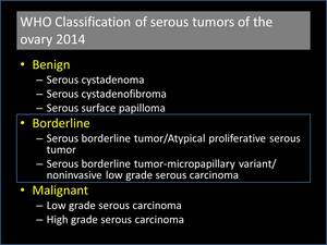
Fig. 1: WHO 2014 classification of serous tumors of the ovary

