ECR 2019 / C-3664
Serous borderline ovarian tumors: tips, tricks and mimics on magnetic resonance imaging
Congress:
ECR 2019
Poster Number:
C-3664
Type:
Educational Exhibit
Keywords:
Genital / Reproductive system female, Oncology, MR, MR-Diffusion/Perfusion, Diagnostic procedure, Multidisciplinary cancer care
Authors:
H. Sahin, Y. Pekcevik; Izmir/TR
DOI:
10.26044/ecr2019/C-3664

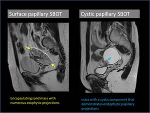
Fig. 2:
Examples of surface and cystic papillary subtypes of serous borderline ovarian...

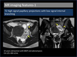
Fig. 3:
T2 high signal papillary projections with low signal internal branching

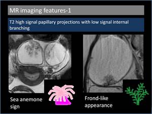
Fig. 4:
Sea anemone sign and frond-like appearance in serous borderline ovarian tumours.

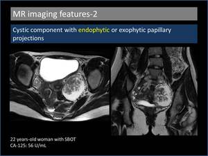
Fig. 5:
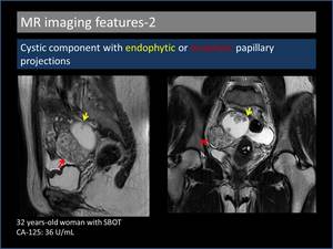
Cystic component with endophytic papillary projections.

Fig. 6:
Cystic component with endophytic and exophytic papillary projections.

Fig. 7:
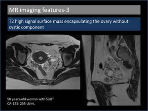
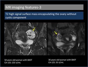
T2 high signal surface mass encapsulating the ovary without cystic component.

Fig. 8:
T2 high signal surface mass encapsulating the ovary without cystic component.

Fig. 9:
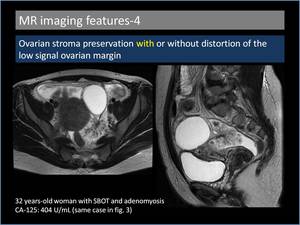
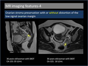
Ovarian stroma preservation with distortion of the low signal ovarian margin.

Fig. 10:
Ovarian stroma preservation without distortion of the low signal ovarian margin

Fig. 11:
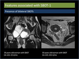
Presence of bilateral serous borderline ovarian tumours.

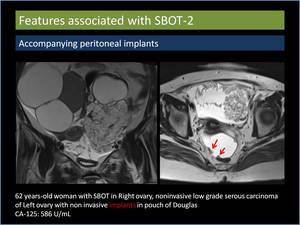
Fig. 13:
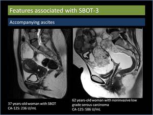
Accompanying ascites.

Fig. 14:
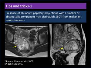
Presence of abundant papillary projections with a smaller or absent solid...

Fig. 15:
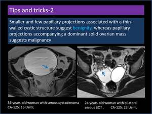
Smaller and few papillary projections associated with a thin-walled cyst

Fig. 16:
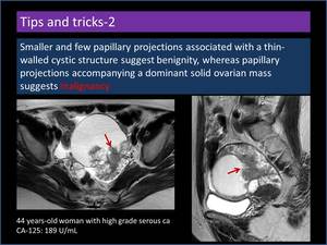
Papillary projections accompanying a dominant solid ovarian mass.

Fig. 17:
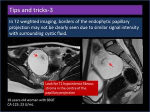
Endophytic papillary projection with subtle borders.

Fig. 18:
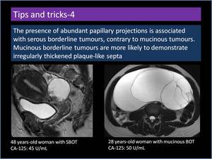
Difference between serous and mucinous borderline tumours.

Fig. 19:
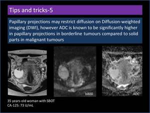
Diffusion-weighted imaging features of papillary projections.

Fig. 20:
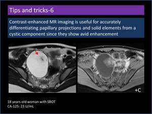
Contrast-enhanced MR imaging features of papillary projections.