Learning objectives
1.To recognize ''never mind'' lesions in paediatric brain MR
2.To understand what are normal variants and incidentalomas
3. To recognize theradiological features that correspond to each normal variantand incidentaloma
4. To understand when do we findnormal variants and incidentalomas and how to deal with each of thempaediatric brain MR
Background
''Never mind'' lesions in paediatric brain imaging usually described as.
Normal variants
Variants of „normal anatomy“
No clinical correlate
No indication for repetition of imaging
Incidental findings (Incidentalomas)
„incidental“ - unexpected – not looked for
Finding as such is abnormal [≠ variant]
Finding does not explain symptomatology
Findings and procedure details
When do we find normal variants and incidentalomas
•„Routine“ – investigations (e.g. following seizure )
•Imaging following trauma
•Neuroimaging in context of studies
(cardiac malformations, follow-up of neonatal HIE…)
•Healthy volunteers («controls» in studies)
•Prenatal screening !!
•Patients with a syndrome – MRI «for interest»
•Check-up „ Wellness scans “
Normal variants
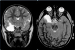
•Temporal lobe hypoplasia–middle fossa arachnoid cyst (Fig.1)
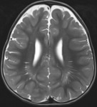
•Enlarged Virchow-Robin spaces ( Perivascular Spaces ) (Fig.2)
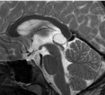
•Pineal cyst (Fig.3)
•Rathke cyst
•Developmental Venous Anomaly [DVA]
•Benign enlargement of subarachnoid space ( frontal / occipital...
Conclusion
Normal variants/ incidental findings are very prevalent
Recognition is relevant
Avoid unnecessary further investigations (and costs)
Avoid (inappropriate) parental anxiety
Personal information and conflict of interest
Marine Grigoryan Gagik
The head of radiology department ''Arabkir'' JMC, Yerevan, Armenia
Associate Professor of Yerevan State Medical University radiology department, Yerevan, Armenia
References
Incidental findings on brain imaging in the general pediatric population , Janses RP et al, NEJM, 2017,377:1593-1595


