Type:
Educational Exhibit
Keywords:
Emergency Imaging, Emergency, Lung, Pulmonary vessels, CT, Digital radiography, Education, Venous access, Blood, Hypertension, Pathology, Not applicable, Performed at one institution
Authors:
S. Stuppner, A. Ruiu; Bolzano/IT
DOI:
10.26044/ecr2020/C-01975
Findings and procedure details
Findings and procedure details
Differentiating HAPE from pneumonia or heart failure can be difficult. Rapid response to oxygen therapy strongly suggests the diagnosis of high-altitude pulmonary edema. Very difficult to diagnose is HAPE with concomitant pneumonia.
Imaging of HAPE
Imaging findings in high altitude pulmonary edema are bilateral multiple prevalent perihilar sided vascular opacities in the lung parenchyma at chest radiograph (Fig 2).
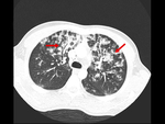
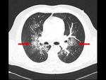
On CT we can see bilateral multiple peribroncovascular parenchymal consolidations, partially confluent (Fig.3 and Fig.4). Pneumonia can have extensive infiltrates on chest radiograph with or without pleural effusion, lobar consolidations, interstitial infiltrates and/or cavitation.
Signs on CT can be from tree in bud signs to ground glass opacities and lobar consolidations. Acute pulmonary embolism shows non-pathognomonic signs on chest radiograph. Acute decompensated heart failure-signs on chest radiograph can be an alveolar edema with bat wing opacities, Kerley B lines, cardiomegaly, dilated upper lobe vessels and pleural effusions. Thickened septal lines due to interstitial edema, ground glass opacity in the dependent part of the lungs and bilateral pleural fluid in CT are characteristic signs.


