Historically, the importance of right ventricle (RV) has been underestimated and overlooked in clinical practice and literature.
Usually, left ventricle (LV) function is most commonly reported.
In recent years, RV function in left heart disease has received more and more attention, the importance of assessment of RV function in clinical management of cardiopulmonary disorders has been recognized .[1-4]
RV dysfunction is an important diagnostic and prognostic indicator in many cardiac and pulmonary diseases. [5-8]
Therefore, to identify RV pathology, guidelines have been published by the American Society of Echocardiography (ASE) on parameters.[9]
Although Echocardiography is usually used as the first diagnostic tool to assess size and function of the right heart, accurate quantitative assessment remains difficult given its complex anatomy.
Echocardiography has some handicaps such as a poor acoustic window and operator dependency.
On the other hand, although CT is not a first diagnostic tool for RV assessment, CT is performed in many cardiac diseases.
As a result of the improvements in CT scanner, because increased spatial and temporal resolution and extended coverage per tube rotation, the continuous acquisition of CT in spiral mode during ECG gating generates a data set that contains all information about the phases of the cardiac cycle.
Also, using the voxel-to-voxel registration motion coherence algorithm that improves the frame rate by filling in the phase-interpolated, the CT can provide a more natural image of cardiac cycle (Fig.1).[13]
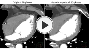
Fig. 1: Comparison between original 10-phase and phase-interpolated 30-phase
However, there are relatively few studies on the evaluation of the right side of the heart with CT, because most studies are conducted with echocardiography or cardiac MRI.
The purpose of this study was to compare 128-slice MDCT and Echocardiography in terms of displaying right ventricular function.
