ECR 2020 / C-04166
Be aware of the epiploic appendagitis
Congress:
ECR 2020
Poster Number:
C-04166
Type:
Educational Exhibit
Keywords:
Not applicable, Inflammation, Education, Ultrasound, MR, CT, Emergency, Anatomy, Abdomen, Emergency Imaging
Authors:
Y. Lamprecht1, R. Pellón1, J. Crespo del Pozo2, E. Marin1, S. Sánchez Bernal1, M. M. Acebo Garcia 1, F. J. GONZALEZ1, E. Montes Figueroa1, J. Azcona Saenz1; 1Santander/ES, 2Selaya, Cantabria, España/ES
DOI:
10.26044/ecr2020/C-04166

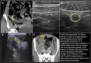
Fig. 2

Fig. 3

Fig. 4

Fig. 5

Fig. 6

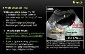
Fig. 7