Type:
Educational Exhibit
Keywords:
Vascular, Arteries / Aorta, Cardiovascular system, Emergency, CT, CT-Angiography, CT-High Resolution, Computer Applications-3D, Computer Applications-Detection, diagnosis, Technical aspects, Acute, Aneurysms, Arteriosclerosis, Not applicable, Observational
Authors:
U. S. Umer, S. Alam, S. Ghulam ghaus, A. Nawaz Khan, S. Gul, H. Abid, N. Gul; Peshawar/PK
DOI:
10.26044/ecr2020/C-05646
Background
Most aortic diseases are associated with atherosclerosis (i.e., aneurysms and dissection), however the spectrum of aortic disease is vast and includes various congenital and acquired entities. Radiologists should be familiar with aortic diseases and with their findings on multidetector CT1. Imaging information that is important to surgeons includes the diagnosis, location of lesion and extent of disease.
Brief Anatomy of Aorta:
Thoracic aorta can be divided into several segments,Root,Ascending aorta,Arch and Descending aorta. Aortic root has three components:Annulus, Sinuses of Valsalva, Sinotubular junction.
The coronary arteries originate from the sinuses adjacent to the sinotubular junction. The ascending aorta extends from the sinotubular junction to the origin of brachiocephalic artery. The arch extends from the brachiocephalic artery origin to the left subclavian artery origin. Its most distal aspect, which is often slightly narrowed, is termed the aortic isthmus. The descending thoracic aorta extends from the ligamentum arteriosum to the level of the diaphragmatic hiatus2. The most proximal portion of the descending thoracic aorta appears slightly dilated and is called the aortic spindle3. Three great arterial branches arise sequentially from the aortic arch.
The abdominal aorta extends from the diaphragm to the level of the fourth lumbar vertebra, where it bifurcates into the right and left common iliac arteries.
Aortic measurements on CTA:
Aortic measurements are made in true short axis projection acquired from double oblique views, from one blood-wall boundary to the other. With CT, ECG gating is essential for accurate measurement, particularly in the ascending aorta (Figure 1).
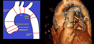
Fig. 1: Measurement of aortic size by CT scan. Double oblique short axis image of mid ascending aorta should be acquired from orthogonal coronal and sagittal images at the level of mid ascending aorta. The thoracic aortic diameters are labelled measured at different levels. 3D image shows markedly increased diameter of aortic root consistent with aneurysm.
Table 1 provides practical values of accepted maximal normal diameters of the aorta1.
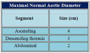
Table 1


