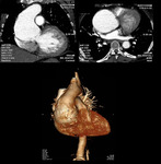
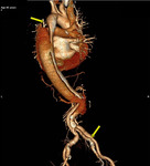
Learning objectives
To evaluate the reliability of 128-slice Multidetector computed tomographic (MDCT) angiography in differentiating different types of aortic diseases and for preoperative aortic morphologic assessment.
Background
Most aortic diseases are associated with atherosclerosis (i.e., aneurysms and dissection), however the spectrum of aortic disease is vast and includes various congenital and acquired entities. Radiologists should be familiar with aortic diseases and with their findingson multidetector CT1. Imaging information that is important to surgeons includes the diagnosis, location of lesion and extent of disease.
Brief Anatomy of Aorta:
Thoracic aorta can be divided into several segments,Root,Ascending aorta,Arch and Descending aorta.Aorticroot has three components:Annulus, Sinuses of Valsalva, Sinotubular junction.
The coronary arteries originate from...
Findings and procedure details
CT Technique:
CTA of the aorta should cover the area from a level 3 cm above the aortic arch to the level of the femoral heads. Currently the standard tube voltage for CTA is 120 kV. The tube current should be approximately 120 mAs, and automated dose modulation should be used. To avoid such artifacts related to beating heart, use of retrospective ECG gating is recommended for imaging of the heart, the aortic root, and the ascending aorta.In this review, images are from CTA performed...
Conclusion
CT is the ideal noninvasive imaging examination for the evaluation of aortic abnormalities. Knowledge of the imaging appearances of aortic lesions enables preoperative or interventional assessment and post-procedural follow up for detection of complications.
Personal information and conflict of interest
Primary author:
Dr. Ummara Siddique Umer
MBBS (KEMC), FCPS (Radiology) , EDiR (EBR)
Consultant Radiologist & Assistant Professor of Radiology
Rehman Medical Institute Peshawar Pakistan
Disclosure:
U. S. Umer; Peshawar/PK - nothing to disclose.
S. Alam; Peshawar/PK - nothing to disclose.
S. Ghulam Ghaus; Peshawar/PK - nothing to disclose.
A. Nawaz Khan; Peshawar/PK - nothing to disclose.
S. Gul; Peshawar/PK - nothing to disclose.
H. Abid; Peshawar/PK - nothing to disclose.
N. Gul; Peshawar/PK - nothing to disclose.
References
Kimura-Hayama ET, Meléndez G, Mendizábal AL, Meave-González A, B. Zambrana GF and Corona-Villalobos CP. Uncommon Congenital and Acquired Aortic Diseases: Role of Multidetector CT Angiography. RadioGraphics .2010;30:1, 79-98.
Rajiah P. CT and MRI in the Evaluation of Thoracic Aortic Diseases. Int J Vasc Med 2013:797189.
Litmanovich D, Bankier AA, Cantin L, Raptopoulos V and Boiselle PM. CT and MRI in Diseases of the Aorta. American Journal of Roentgenology .2009; 193:4 , 928-40.
Bosniak MA. An analysis of some anatomic–roentgenologic aspects of the brachiocephalic vessels. Am...


