According to ACR BI-RADS 5th Edition 2013, non-mass enhancement (NME) is defined as a discrete enhancing area that does not represent a mass. It typically interspersed with non-enhancing fatty or glandular tissue. NMEs can be either benign or malignant. It is most commonly due to fibrocystic change including focal adenosis, hormonal stimulation, inflammatory changes, or intraductal and diffuse cancer. Ductal carcinoma-in-situ (DCIS) have been reported as the most common manifestation of NMEs. Hence, pattern recognition of malignant NMEs are essential.
According to ACR BI-RADS, NMEs are described based on their distribution and internal enhancement pattern ( Fig. 1 ).
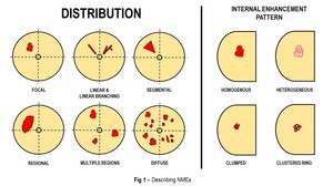
Fig. 1: Describing NMEs
Distribution
a. Focal: Refers to confined area of enhancement which is less than a breast quadrant volume.
b. Linear & Linear Branching: Refers to enhancement arrayed in line or a line that branches following breast duct.
c. Segmental: Refers to a triangular or wedge-shaped enhancement with its apex pointing towards the nipple.
d. Regional: Refers to enhancement that involves a large area of breast tissue, at least a quadrant.
e. Multiple regions: Refers to enhancement in at least two large volume of tissue not conforming to ductal distribution and separated by normal tissue.
f. Diffuse: Refers to enhancement distributed throughout the breast in random fashion.
Internal enhancement pattern
a. Homogenous: Refers to uniform enhancement.
b. Heterogenous: Refers to non-uniform enhancement.
c. Clumped: Refers to cobblestone enhancement of varying sizes and shapes resembling a bunch of grapes or a string of pearls.
d. Clustered Rings: Refers to clustered group of thin rings of enhancement.
With the emerging and increasing use of Breast MRI as a screening tool for breast cancer, the importance for pattern recognition of NMEs cannot be understated. A breast cancer may present only as NME without other significant mammographic findings ( Fig. 2 ). For example, DCIS have been described to exhibit as NMEs in the range of 69 – 90%.
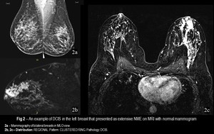
Fig. 2: An example of DCIS in the left breast that presented as extensive NME on MRI with normal mammogram
Authors have identified linear branching distribution as well as clustered rings enhancements as predictors of malignancy. In this exhibit we have provided pictorial review of the different appearances of NMEs and their pathological correlation. Knowledge of the common presentation for both benign and malignant causes of NMEs are crucial for avoiding false negative results.


