Type:
Educational Exhibit
Keywords:
Not applicable, Toxicity, Metabolic disorders, Drugs / Reactions, Imaging sequences, MR, Neuroradiology brain, CNS, Neuro
Authors:
P. Guadalupi1, L. milonia1, A. Marrazzo1, C. Giordano2, F. Magnani3, A. Botto2, T. Tartaglione1, S. Gaudino2, C. Colosimo2; 1Roma/IT, 2Rome/IT, 3Roma, Ragusa (RG)/IT
DOI:
10.26044/ecr2020/C-11893
Background
The brain represents only 2% of body weight, but it consumes about 20% of blood oxygen and 25% of glucose. Due to its high metabolic demands the brain is very vulnerable to toxic substances or metabolic disorders.
Toxic\metabolic encephalopathies commonly present a symmetrical brain involvement, they mainly affect the basal ganglia (BG), the white matter (WM), thalami and cortex(Tab.1).
Gray matter (GM) is highly metabolically active; BG present rich vascularization, high levels of mitochondria and neurotransmitters, globi pallidi (GP) and putamen are particularly susceptible to hypoxia and anoxia; WM is more susceptible to lipophilic toxic substances. The corpus callosum (CC) (the splenium in particular) is vulnerable to cytokinopathy: its neurons, astrocytes, oligodendrocytes have a higher density of receptors (cytokine, glutamate, excitatory-amino-acid receptors and other toxin\drug receptors) that leads to a higher tendency for cytotoxic\excitotoxic edema. Oligodendrocytes of the pons are more vulnerable to osmotic changes.
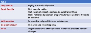
Table 1
There are at least four types of cerebral edema explained by different physiopathological mechanisms and with their own imaging findings: vasogenic, cytotoxic, excitotoxic and intramyelinic edema. They are variously related to toxic and metabolic alterations and their reversibility\irreversibility(Tab.2).
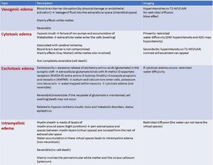
Table 2
Normal MRI-signal:
T1 Weighted-Images(WI):
- Caudate Nucleus (CN), putamina and thalami: isointense with cortex
- GP: vary in signal intensity, physiologic and age-related site of calcification and iron deposition
- WM in internal and external capsules: hypertense relative to the BG
T2-WI:
- CN, putamina, thalami: isointense with cortex
- GP: more hypointense (higher myelin content) relative to the putamen
- “Dark putamen”: normal by 70-80years (increasing iron deposits)
SWI\T2*(echo-gradient-WI):
- GP: hypointense relative to cortex,
- Putamina: iron deposits bloom and (lateral) putamen appears hypointense relative to thalami but not as much as GP


