ECR 2020 / C-13566
Top signs in gastrointestinal radiology
Congress:
ECR 2020
Poster Number:
C-13566
Type:
Educational Exhibit
Keywords:
Not applicable, Education and training, Education, Conventional radiography, Abdomen, GI Tract
Authors:
K. Saravanan1, B. Ashraf ahmed2, E. Premgowtham3, F. Abubacker Sulaiman4; 1CHENNAI, TAMIL NADU/IN, 2Melmaruvathur, Tamil Nadu/IN, 3Chennai/IN, 4Chennai, TN/IN
DOI:
10.26044/ecr2020/C-13566

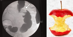
Fig. 1:
Apple core sign- Contrast study showing the appearance of an annular...

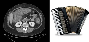
Fig. 2:
Accordion sign- Markedly thickened bowel wall with oral contrast trapped...
:22")
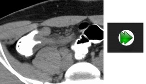
Fig. 3:
Arrowhead sign- Arrowhead shaped inflammatory changes of the cecal base...
 40: 3338.")
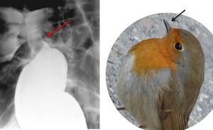
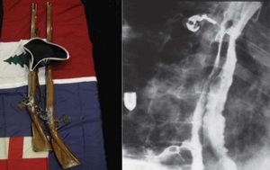
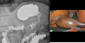
Fig. 4:
Bird’s Beak sign - contrast study showing appearance of achalasia like a...

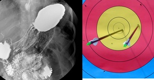
Fig. 5:
Bull's eye sign- A small pit of barium contained within an ulcer cavity in the...
:615-7.")
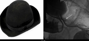
Fig. 6:
Bowler hat sign- Double contrast study showing cup-shaped filling defects that...
 43: 3536.")
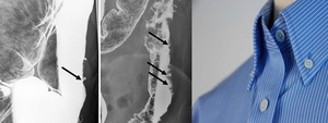
Fig. 7:
Collar button sign- Contrast barium enema shows mucosal ulceration with...
 43: 3532.")
Fig. 8:
Cobblestone sign- Small bowel follow through shows scattered islands of normal...

Fig. 9:
Comb sign- CECT image shows engorged vasa recta secondary to hyperemia of the...
:22")
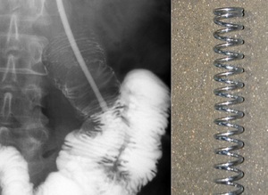
Fig. 10:
Coiled spring sign- Plain radiograph of the abdomen following the...

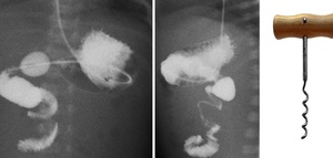
Fig. 11:
Corkscrew sign- describes the spiral appearance of the distal duodenum and...
:22")
Fig. 12:
Double-barrel esophagus- Contrast study demonstrates dissection of oral...

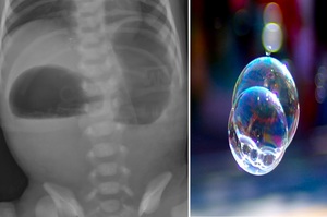
Fig. 13:
Double Bubble Sign- abdominal radiography showing two air-filled structures in...
:22")
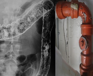
Fig. 14:
Lead pipe sign— Abdominal X-ray showing narrowed and ahaustral segment of the...

Fig. 15:
Leather bottle stomach- Radiograph of the stomach following oral barium...
:22")
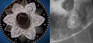
Fig. 16:
Mexican hat sign- Radiograph of an upper GI demonstrates 2 concentric rings....
:22")
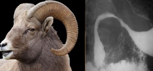
Fig. 17:
Ram's horn sign- Radiograph of the stomach following oral contrast shows...
:22")
Fig. 18:
Ribbon sign- Marked luminal narrowing and effacement of the valvulae...
:22")
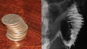
Fig. 19:
Stack of coins- Radiograph of the abdomen following oral contrast represent...
:22")
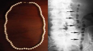
Fig. 20:
String of pearls- Left lateral decubitus radiograph of the abdomen shows a row...

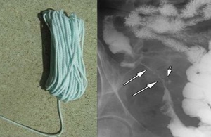
Fig. 21:
String sign- A thin line of barium is seen in the terminal ileum and also small...

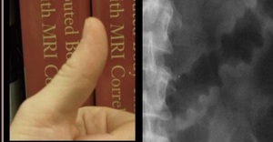
Fig. 22:
Thumbprint sign- Thickening of the haustra secondary to edema and hemorrhage...

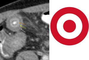
Fig. 23:
Target sign- Hyperdense ring represents the hyperemic mucosa and muscularis...
:22")
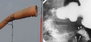
Fig. 24:
Windsock sign- Intraluminal duodenal diverticulum surrounded by narrow...

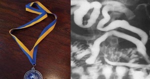
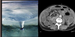
Fig. 25:
Whirlpool sign- superior mesenteric artery and vein wrapping around one another...