Typical and atypical soft tissue tumours of fingers are analysed through different X-ray, US, CT and MRI studies performed at our Institution.
The standard of reference is cytology/histopathology in most of the cases.
- Cystic lesions [1,3,5,7] (Fig. 8)
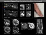
- Ganglion/synovial cyst (Fig. 9)
Both synovial and ganglion cysts have a thin connective capsule and contain fluid or mucinous material so are indistinguishable on imaging. However, the histopathology is distinct, first has synovial lining whereas ganglion has not.
The aetiology is unclear but they may originate from a synovial coalescence of degenerative cyst arising from tendon sheath, joint capsule or bursae.
Its main location is the wrist but can appear in fingers adjacent to:
- The flexor tendon sheath (10%) at the metacarpophalangeal joint secondary to repetitive strain injuries.
- The dorsum of the distal interphalangeal joint (10%) associated with osteoarthritis.
These cysts are filled with keratin debris and lined by a stratified squamous epithelium. Fewer than 10% occur in the extremities.[1]
They are thought to occur as a result of traumatic implantation, congenital rests of epithelial cells or pilosebaceous unit occlusion.
Differential diagnosis includes:
- Ganglia when located near joints.
- Sarcomas and tendon sheath tumours if there is a heterogeneous signal, but these will frequently have internal enhancement.
- Intraosseous epidermoid cyst of the distal phalanx [8](Fig. 12)
Intraosseous epidermoid cysts are exceedingly rare and usually involve the phalanges (most common site the distal phalanx), skull and toes.
Traumatic origin as implantation of epidermal or nail bed fragments into the phalanx remains its most prevalent pathogenesis hypothesis.
Differential diagnosis of osteolytic lesions located at the distal phalanx include:
- Enchondroma
- Glomus tumour
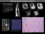
- Giant cell tumour of the tendon sheath
- Less common: bone metastasis, osteoid osteoma, aneurysmal bone cyst
GCTTS are common benign synovial lesions with a predominance of macrophage-related components of unknown aetiology that arise from the tendon sheath.
These lesions usually affect the volar aspect of the first three digits.
- Giant cell tumour of bone (Fig. 19)
Classically benign tumours that arise from the epiphysis of the bone and are locally aggressive.
These tumours are rare benign tumours arising from the tendon sheath that typically present fibrous tissue (myofibroblasts) resulting in low signal on both T1-w and T2-w images.
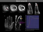
Glomic tumours are hamartomas of the neuro-myoarterial apparatus within the glomus body and account for up to 5% of soft tissue tumours of the hand.
They are commonly found at the fingertip (65% of cases); either in the pulp or beneath the fingernail.
Temperature sensitivity is a typical clinical finding.
Leiomyomas are benign smooth muscle tumours. The typical location is the uterus but they can also be seen in the subcutaneous tissue.
- Peripheral Nerve Sheath Tumours (PNST) (Fig. 26)
Benign PNST includes both schwannomas and neurofibromas, both present in young adulthood as solitary slow-growing lesions.
In the hand and wrist, schwannomas arise from deeper and larger nerves (particularly the ulnar nerve along with the flexor aspects) whereas neurofibromas tend to involve smaller cutaneous nerves.
Another characteristic that may allow differentiation is that schwannomas tend to be encapsulated and eccentric to the nerve whereas neurofibromas arise from the central nerve fascicles.
Benign and 10% of malignant PNSTs may have a large myxoid component.
Malignant soft tissue tumours of the fingers are uncommon.
PFT is a rare mesenchymal neoplasm primarily occurring in young adults, classified as intermediate malignancy because of the local aggressivity, recurrence and possibility of lymph node and distant metastases.
The most common location is the upper extremities.
Primary surgical excision to achieve remains the treatment of choice.
Soft tissue metastases are very rare and are often related to widespread disease or locoregional recurrence.
Imaging features are similar to that of soft tissue primary malignancies.
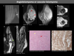
Haemangiomas are low-flow benign vascular tumours that consist of cellular proliferation and hyperplasia.
They are characterized by a rapid proliferative stage and later involution phase (6-10 months) and typically appear in the neonatal period but some can persist into adulthood (may present incidentally or after trauma).
Vascular malformations arise from dysplastic vascular channels and exhibit a normal rate of endothelial turnover, growing commensurately without regression. They can enlarge due to haemorrhage, infection or hormonal influence.
The incidence in fingers is rare. They are usually confined to the subcutaneous tissues but can have an intraarticular or intraosseous extension.
- Low-flow: Venous malformation (Fig. 34)
- High-flow
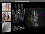
Arteriovenous malformations are typically congenital and consist of feeding arteries, draining veins and a nidus composed of multiple dysplastic vascular channels with the absence of a normal capillary bed.
Arteriovenous fistulas are acquired and formed by a single vascular channel between an artery and a vein. (Fig. 35,Fig. 36,Fig. 37, Fig. 38, Fig. 39)
- Other: Infectious, Inflammatory, Traumatic
Soft tissue infection with abscess formation may mimic a mass.
MR allows demonstration of the extent and involvement of soft tissue and helps to determine the presence of osteomyelitis.
Gout arthropathy is due to deposition of monosodium urate crystals in soft tissues and joints secondary to hyperuricemia states.
Characteristic imaging findings include juxta-articular erosions and calcified dense soft tissue nodes.
Gouty tophi are foreign body granulomas that include macrophages and crystals and can present as focal masses.
(Fig. 42)
Psoriatic arthritis is one of the seronegative spondyloarthropahy-spectrum diseases related to psoriasis.
It typically has a nail and distal interphalangeal joint involvement with asymmetrical and oligoarticular affectation (this allows differentiation with rheumatoid arthritis).
Synovial inflammation and bony erosions can simulate infectious conditions.(Fig. 43)
These are benign fibro-adipose subcutaneous pads located para-articular over the dorsal aspect of proximal interphalangeal joints.
Various types of conditions can affect the subungual space.
Traumatic injuries of the extensor and flexor tendons can cause soft tissue lesions in the fingers.


