ECR 2020 / C-14393
Tuberculosis: can AI become a reliable method for diagnosis and screening?
Congress:
ECR 2020
Poster Number:
C-14393
Type:
Educational Exhibit
Keywords:
Artificial Intelligence and Machine Learning, Artificial Intelligence, Computer applications, Thorax, Conventional radiography, Teleradiology, Computer Applications-Detection, diagnosis, Computer Applications-General, Computer Applications-Teleradiology, Cavitation, Image verification, Outcomes, Retrospective, Diagnostic or prognostic study, Performed at one institution
Authors:
A.-E. Moldovan, C. Avramescu, B. Bercean, S. Iarca, A. Tenescu, F. Birsasteanu; Timisoara/RO
DOI:
10.26044/ecr2020/C-14393

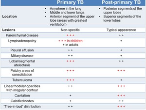
Fig. 1:
Radiographic features of primary and post-primary TB [4].