ECR 2020 / C-14718
Structured Reporting Format for CT and MR liver dynamics for hepatocellular carcinoma: Is it worth doing?
Congress:
ECR 2020
Poster Number:
C-14718
Type:
Educational Exhibit
Keywords:
Performed at one institution, Not applicable, Quality assurance, Neoplasia, Education and training, Imaging sequences, History, Education, MR-Diffusion/Perfusion, MR, CT, Biliary Tract / Gallbladder, Anatomy, Abdomen, Abdominal Viscera
Authors:
A. N. Khan1, K. S. Babar2, U. S. Umer2, W. Farman2, N. Safi2; 1Leicetser/UK, 2Peshawar/PK
DOI:
10.26044/ecr2020/C-14718

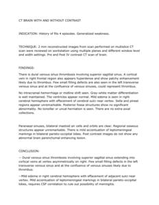
Fig. 2:
Example of first level reporting format.

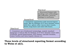
Fig. 1:
Three levels of structured reporting format.

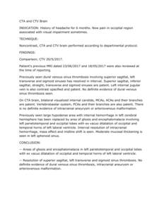
Fig. 3:
Example of second level reporting format.

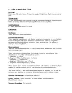
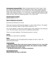
Fig. 4:
Example of third level reporting format page 1.

Fig. 5:
Example of third level reporting format page 2.