ECR 2020 / C-15049
The role of Magnetic Resonance Imaging in the assessment of carotid space masses
Congress:
ECR 2020
Poster Number:
C-15049
Type:
Educational Exhibit
Keywords:
MR, Contrast agent-intravenous, Neoplasia, Education and training, Head and Neck
Authors:
O. Medvedev, L. M. Lenghel; Cluj-Napoca/RO
DOI:
10.26044/ecr2020/C-15049
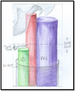
 as a tube extending from skull base to the aortic arch. The hyoid bone divides the CS into suprahyoid (SHN) and infrahyoid (IHN) portion. VS – visceral space, PVS – perivertebral space, SL-DCF - supeficial layer of deep cervical fascia.
References: Medvedev Olga; adapted from [1]")
Fig. 1:
Lateral graphic of the neck depicting the carotid space (CS) as a tube...

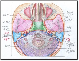
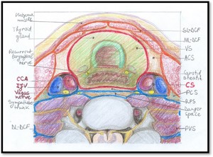
Fig. 2:
Axial graphic of the SHN at the level of C1 vertebral body. MS – masticator...

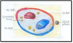
Fig. 3:
Graphic illustration of magnified left SHN CS. The carotid artery lies in the...
. At this level a CS mass will displace the parapharyngeal space anteriorly and the styloid process anterolaterally.
References: Emergency County Hospital Cluj – Napoca, Romania")
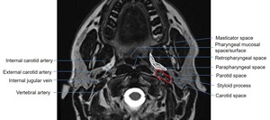
Fig. 4:
Axial T2 image at the level of C1 vertebral body. The SHN CS contains the...

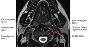
Fig. 5:
Axial T2 image at the level of mid-oropharynx. The posterior belly of the...

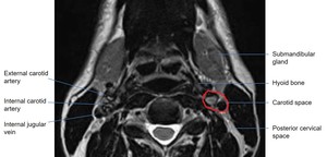
Fig. 6:
Axial T2 image at the level of hyoid bone. At this level the carotid...

Fig. 7:
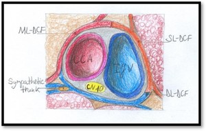
Axial graphic of the CS in the IHN. The carotid sheath contains all 3 layers of...
, internal jugular vein (IJV) and only the vagus cranial nerve (CN10). SL-, ML-, DL-DCF – superficial, middle and deep layer of deep cervical fascia
References: Medvedev Olga, magnified from fig. 7")
Fig. 8:
Graphic illustration of magnified left IHN CS. Note that at this level the CS...

Fig. 9:
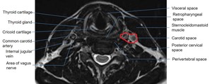
Axial T2 image at the level of cricoid cartilage. Surrounding tissue...

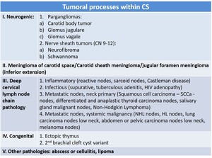
Table 1:
Tumoral processes that can occur within the carotid space. CN – cranial...
 and examples of clinical implications of CS masses (adapted according to [1]). SCCa - Squamous cell carcinoma; CN – cranial nerves; CA- carotid artery; IJV – internal jugular vein.
References: adapted according to [1, 4]")
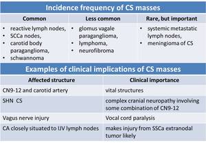
Table 2:
Incidence frequency of CS masses (adapted according to [1, 4]) and examples of...