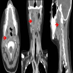
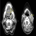
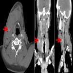
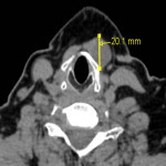
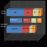
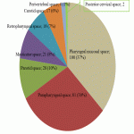
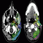
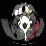
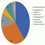
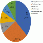
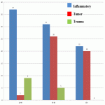
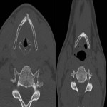
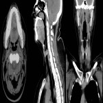
Neck soft tissue pathology was found in 175/208 cases: 90 cases inflammatory changes (Fig.2.), 48 cases oncology (Fig.3.), 14 cases trauma (Fig.4.) and 18 cases other pathology, for example congenital pathology (Fig.5.,6.). Most cases showed involvement above os hyoideum - pharyngeal space (n=100), parapharyngeal space (n=81), parotid (n=28), masticator (n=21), less often retropharyngeal (n=18), carotid, (n=17), perivertebral (n=4) and posterior cervical space (n=2) (Fig.7.,8.,9)
Most commonly inflammation and abscess involved pharyngeal mucosal space, parapharyngeal space, parotid space, masticator space similar to tumor (Fig.10.,11.).
In case of inflammation and abscess superficial soft tissues were involved in 11 cases, visceral space in 8 cases. Tumor involved superficial soft tissues 4 cases, visceral space 12 cases. In case of a trauma most commonly visceral space was involved (n=7), less often parapharyngeal space (n=3), and pharyngeal mucosal space, parotid space, masticator space, retropharyngeal space, carotid space accordingly in two cases.
Pharyngeal and parapharyngeal space involvement was overlapping in oncology and inflammatory cases (39% vs 34%; 34% vs 27%), respectively (p>0.05). From 54 histologically confirmed tumors, most often laryngeal tumor (n=15), palatine tonsil (n=10), tongue root tumor (n=9), pharyngeal tumors (n=7), non-Hodgkin lymphoma (n=3) were detected, sparsely metastatic tumors without knowledge of primary tumor localization, salivary gland tumors. Inflammatory changes were seen in all age groups, tumors >45y, trauma predominantly <44y (p>0.05). CRP threshold >125 mg/l was suggestive for abscess (p>0.001). Enlarged lymph nodes were non-specific in 90/108 cases, enhancement >70HU; specific (Fig.3.) with structural changes and heterogeneous contrast enhancement in 18/108 cases (confirmed histologically). Bilateral lymph node involvement in 57%; predominantly in the groups I(n=53), II(n=80), III(n=41), less often IV (n=11), V (n=2), VI (n=6) groups.
With age related groups inflammatory changes in neck soft tissues were relatively similar, but tumor more often affects elderly patients after 45 years, it was relatively rare in age group from 18 till 44 years.
Emergency medical assistance in case of a trauma most often was in the age group from 18 till 44 years (n=21), less often from 45 till 64 years of age (n=8), and in one case after 65 years of age, where CT was without pathological finding (Fig.12).
From 105 patients with inflammatory changes in 64 cases CT was found, from which 25 patients were treated surgically, with local anesthesia and incision, drainage of abscess. In five cases surgical treatment was performed with aim to evacuate parotid salivary gland duct stones.
Histology and less frequently performed cytology (n=12) examination helps differentiate inflammatory changes from oncologic changes. Cytology examination in nine cases revealed purulent content, in two cases malignant cells and in one case there was cytology smear test without the suspicion of oncology and further histology examination was recommended.
Youngest patient with tongue cancer was 31 years old, average patient age with oncological diseases was 52.3 years (median 52 years), oldest patient was 93 years old.
In five cases patients with a history of oncological diseases and clinical complaints about hemoptysis had been recommended intervention radiologist consultation, additional, digital subtraction angiography (DSA) and peripheral embolization was performed with the occlusion of artery with extravasation, for example, for a patient with palatine tonsill malignant tumor external carotid artery branch endovascular embolization with microspheres and embolization coils were used to stop the bleeding.
Discussion
CT examination is performed for inflammatory, tumor created changes (also when there is a suspicion of extravasation from it), in a trauma and other cases where acute changes can be combined (for example an old fracture (Fig.13). If a patient was treated for example, with radiation therapy, surgical therapy, lymphnode extirpation etc. and if data in the emergency stage refers to known oncology history, for example, in the chest or abdomen, then it is easier to narrow the radiological differential diagnosis.
Comparing the data from the literature with the results from our studies above os hyoideum most commonly pharyngeal mucosal space was involved in the pathological changes, following parapharyngeal space, parotid space which showed similar results from the literature, although there are controversial data found as well, where, from 60 patients with clinical suspicion for pathological changes in neck ,most commonly (n=28) it was a tumor and the inflammatory changes were the second most common reason (1). In this study the most common changes were due to inflammatory, less common data was obtained about oncology or congenital neck lesion.
In CT examination with a suspicion of inflammatory changes, including abscess, where it was as a collection of low density fluid with enhancing walls (Fig.2.), abscess was determined in most (85.7%) cases. Most common interfascial space with diagnosis of abscess was in pharyngeal mucosal space, second most common parapharyngeal space similarly to the previous study (2).
In cases where there is a suspicion about oncology, in literature (3) it is suggested to perform examination in two contrast enhanced series, which in return could give additional information about neoprocess, and sometimes tumor or lymph nodes could enhance earlier or later if the examination is performed only at 100th second as one post-contrast phase. Additional phase prolongs examination time and a patient with a tumor still needs additional examination, for example, magnetic resonance imaging, which then allows to evaluate the soft neck tissues more detailed. Neck soft tissue evaluation with CT with contrast-enhanced 3-series could help better differentiate pathological changes (e.g. oncology), however, to reduce the radiation dose for the patient, examination at the 100th second after contrast injection could be optimal to evaluate soft tissues and there would be enough contrast enhancement in the vessels to characterize them. For optimal examination of the arteries and veins enhancement this may be sufficient, although for neck tumors, lymph nodes evaluation it may take a longer time (sometimes even 180th seconds) to enhance.

