Keywords:
Head and neck, Ultrasound, Contrast agent-intravenous, Localisation, Endocrine disorders, Neoplasia
Authors:
S. Pavlovics, M. Radzina, Z. Narbuts, M. Miķelsone, M. Ratniece, R. Niciporuka, M. Liepa, P. Prieditis, A. Ozolins
DOI:
10.26044/ecr2022/C-10658
Methods and materials
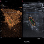
Prospective study included 75 patients (18-77 years, F:M=64:11, with hyperparathyroidism scheduled for parathyroidectomy. B-mode ultrasound, Colour Doppler, Superb Microvascular Imaging(SMI), CEUS (SonoVue 1 ml vs. 2ml) images obtained and postprocessing performed. Results compared with postoperative morphology - 68 parathyroid adenomas (PA), 17 parathyroid hyperplasia (PH). It may be challenging to differentiate parathyroid lesions from lymph nodes and thyroid nodules in B-mode US because of visual similarities between these structures, especially in patients with goitres and after previous neck surgery [1-3]. Contrast-enhanced US with intravenous administration of a contrast agent, is being advised to improve the accuracy of conentional US [1,4]. The European Federation of Societies for Ultrasound in Medicine and Biology (EFSUMB) approved the use of CEUS as a safe method for both children and adults [5,6]. The mechanical index was reduced to less than 0.10 during CEUS examination, and the B-mode US and CEUS parallel imaging was obtained. Quantitative evaluation of acquired raw CEUS data using VueBox (Bracco, Italy) application was performed in 51 lesions (5 hyperplasias, 46 adenomas). Regions of interest (ROI) in the central part (green) of parathyroid lesions and periphery (orange) were created. Wash-in rate (WiR), Wash-out rate (WoR) and Average Contrast Signal Intensity (ACSI) of selected ROI were analysed during quantitative analysis.


